

ABSTRACT
Introduction: Takayasu arteritis is the most common large-vessel vasculitis in childhood, but there is a lack of literature regarding childhood-onset Takayasu arteritis (c-TAK) in Southeast Asia. We aim to describe a c-TAK cohort in Singapore and highlight a unique subset that first presents with Kawasaki-like disease (KD).
Method: A single-centre cohort study in Singapore of consecutive children diagnosed with c-TAK between 2002 and 2023 was performed. Demographic and clinical features, laboratory and angiographic findings, treatment, and outcomes were summarised. Disease activity was evaluated using the Paediatric Vasculitis Disease Activity Score and inflammatory markers.
Results: Twenty-three patients, fulfilling both the EULAR/PRINTO/PReS and ACR/EULAR 2022 criteria, were recruited. The most common clinical features at diagnosis were fever (15, 65%) and neurological symptoms (11, 48%, half of which presented with stroke), while the most prevalent angiographic pattern by Hata’s classification was Type V (21, 91%). Eight children (35%) initially presented as refractory KD, and these patients were significantly younger, more male-predominant, and had higher inflammatory markers at diagnosis; all of them had coronary artery involvement, but none had intracranial vascular findings. Of the entire cohort, 16 (70%) achieved inactive disease on medications with a median duration of 6 months (interquartile range [IQR]: 4–11), and 8 (35%) achieved remission off medications with a median duration of 43 months (IQR 35–60).
Conclusion: Our c-TAK cohort has high proportions of neurological involvement and stroke. This is also the first cohort study to describe a distinct group of patients who first presented with refractory KD.
CLINICAL IMPACT
What is New
- This is the first reported cohort of childhood-onset Takayasu arteritis (c-TAK) in Southeast Asia, showing unique characteristics including high proportion of central nervous system involvement.
- To the authors’ knowledge, this study is the first to describe a distinct subgroup of c-TAK patients who initially presented with Kawasaki disease.
Clinical Implications
- Findings highlight the need to screen for large-vessel vasculitis in patients presenting with young-stroke or refractory Kawasaki disease.

Takayasu arteritis (TAK) is the most common large-vessel vasculitis in childhood. It is characterised by granulomatous inflammation of the aorta and its major branches, resulting in segmental stenosis, occlusion, dilatation, and/or aneurysms.1 Compared to adult-onset TAK, childhood-onset Takayasu arteritis (c-TAK) presents with more systemic and constitutional features, has distinct vascular involvement, and is often a more refractory disease that requires greater immunosuppression.2 Separate classification criteria have also been proposed for c-TAK after a joint effort between the European League Against Rheumatism (EULAR), the Paediatric Rheumatology International Trials Organisation (PRINTO), and the Paediatric Rheumatology European Society (PReS).3 Given the nonspecific systemic symptoms at initial presentation, the diagnosis of c-TAK is often difficult.4,5
Kawasaki disease (KD), on the other hand, is a vasculitis that preferentially affects medium-sized arteries, particularly the coronary arteries (CA).6 As a result of the strong association between KD and CA aneurysms, febrile children with CA dilation or aneurysms are almost always diagnosed with KD. However, there have been case reports of children who were initially managed as KD and were later diagnosed with c-TAK.7-10
Notably, there is considerable variation in the incidence and characteristics of adult TAK cohorts across different geographic regions.11,12 Such epidemiological variability is also mirrored in c-TAK cohorts around the globe.13-23 However, there is a paucity of literature regarding c-TAK cohorts in Southeast Asia. To address these issues, we aim to describe the clinical characteristics, radiological findings, and outcomes of our c-TAK cohort, and compare them with cohorts in other geographic regions. We also aim to describe a subset of patients with KD as the initial presentation and examine any differences in the clinical course compared with other c-TAK patients.
METHOD
Study population
Patients with c-TAK were recruited at the KK Women’s and Children’s Hospital, Singapore, from August 2002 to January 2023. The institution is the only children’s hospital in Singapore and one of only 2 paediatric rheumatology centres nationwide, managing two-thirds or more of all referral cases. All patients were younger than 18 years at onset and were diagnosed by trained paediatric rheumatologists. The diagnosis was established based on clinical findings and angiographic images compatible with the disease, excluding other possible causes. All recruited patients fulfilled both the 2010 EULAR/PRINTO/PReS classification criteria and the 2022 American College of Rheumatology (ACR)/EULAR classification criteria.3,24
Data collection
This retrospective cohort study utilised data from our prospective registry (Registry for Childhood-Onset Rheumatic Diseases).25 Demographic, clinical, laboratory tests, imaging studies and treatment data were prospectively collected at diagnosis and during each follow-up visit following a standardised protocol. Arterial hypertension was defined as blood pressure >95th percentile for age and height. Blood pressure discrepancy was defined as a difference of ≥10 mmHg between limbs. Antinuclear antibody (ANA) was determined by indirect immunofluorescence on human cell epithelioma (Hep-2) cells substrate and defined as positive if staining reactivity was ≥1:160. Antineutrophil cytoplasmic antibodies were determined using indirect immunofluorescence. Tuberculosis screening was done through both tuberculin skin tests (positive test defined as ≥10 mm or more regardless of age)26 and the interferon-gamma release assay (IGRA). Vascular imaging studies were performed at the discretion of attending physicians, which included computed tomography angiography, magnetic resonance angiography (MRA), fluorodeoxyglucose (FDG)-positron emission tomography (PET) scan, and echocardiography studies. An arterial segment was considered affected if it demonstrates one or more of the following radiological changes: mural enhancement, thickening, or irregularity, stenosis, occlusion, dilatation, or aneurysm. The territorial distribution of lesions as defined by Hata’s angiographic classification was recorded.27 No patients were excluded due to incomplete data.
Outcomes
Disease activity was assessed at each visit using the validated Paediatric Vasculitis Activity Score (PVAS). PVAS is currently the only validated disease activity tool specific to paediatric vasculitis and is used as an outcome measure in paediatric vasculitis clinical trials.28 Active disease was defined as PVAS ≥1 and/or raised inflammatory markers and/or imaging evidence of active disease, including new angiographic lesions and vessel wall inflammation by post-contrast enhancement and thickening. Partial remission was defined as a PVAS score of 0, erythrocyte sedimentation rate (ESR) <40mm/h, C-reactive protein (CRP) <20 mg/L, and prednisolone dose ≤0.15 mg/kg/day or ≤7.5 mg/day, whichever is lower.29-31 Complete remission (CR) was defined as a PVAS score of 0, ESR <20 mm/h, CRP <10 mg/L, and prednisolone dose ≤0.15 mg/kg/day or ≤ 7.5 mg/day, whichever is lower.29-31 Clinically inactive disease on medication (CID) is defined as CR while on glucocorticoids or disease-modifying anti-rheumatic drugs (DMARDs), while clinical remission off medication (CRM) was defined as CR off all medications. Disease flare was defined as the occurrence of new c-TAK symptoms and/or an increase in inflammatory markers necessitating altered treatment and/or new or worsening angiographic lesions.32
Statistical analyses
Non-parametric analyses were used to describe data and were shown as median (25th–75th percentiles) for continuous variables and percentages for categorical variables. Chi-squared or Fisher’s Exact, Mann-Whitney U, or Kruskal-Wallis tests were applied to compare groups where appropriate. A Kaplan-Meier survival curve was constructed for median time to outcomes. All analyses were performed using Stata 18 (StataCorp, College Station, TX, US) and GraphPad Prism v7 (GraphPad Software, CA, US) with statistical significance set at P<0.05.
Ethics
This study was approved by the SingHealth Centralised Institutional Review Board (CIRB 2019/2961 and 2019/2194). Informed consent was waived due to the nature of the retrospective data review from the registry.
RESULTS
Demographic features and clinical characteristics
A total of 23 patients with c-TAK (10 females [44%], 13 Chinese [57%]) were included. The median age at diagnosis was 3.9 (0.6–11.9) years, and the median duration of follow-up was 4.0 (0.7–6.1) years (Table 1). Eight patients (35%) initially presented with clinical features of KD and were managed as such. Seven of them fulfilled the 2017 American Heart Association Diagnostic Criteria for KD.33 However, their disease was refractory to the first and second-line treatments (either a second dose of intravenous immunoglobulin [2 patients] or intravenous pulse methylprednisolone [4 patients], or both [2 patients]), which prompted further evaluation and eventually diagnosis of large-vessel vasculitis (referred to as the “c-TAK-KD” group herein). The median age of diagnosis of this subgroup was younger at 0.5 (0.4–1.8) years compared to the typical c-TAK group (11.4 [3.9–13.5] years, P<0.001). Individual characteristics of these 8 c-TAK-KD patients are summarised in Supplementary Table S3.
Table 1. Demographic, clinical and laboratory features at diagnosis.
Overall, the most common presenting clinical features at diagnosis were fever (15 patients, 65%), neurological symptoms (11 patients, 48%), hypertension (9 patients, 39%) and gastrointestinal symptoms (38 patients, 5%, Table 1). Compared to the typical c-TAK group, the c-TAK-KD group had much higher proportion of patients with fever (8 patients [100%] versus [vs] 7 patients [47%], P<0.001), conjunctival injection (5 patients [63%] vs 1 patient [7%], P=0.009), mucosal changes (5 patients [63%] vs 0 patient [0%], P=0.002), cervical lymphadenopathy (3 patients [38%] vs 0 patient [0%], P=0.032) and extremity changes including desquamation, oedema and palmar erythema (4 patients [50%] vs 0 patient [0%], P=0.008). Patients in the typical c-TAK group had more neurological symptoms (10 patients [67%] vs 1 patient [13%], P=0.027), of which a significant proportion presented with headache (6 patients, 40%), altered sensorium (5 patients, 33%) and stroke (5 patients, 33%).
Laboratory findings
Patients in c-TAK-KD group presented with lower haemoglobin levels (9.6 [8.9–10.7] g/dL vs 12.6 [10.9–13.3] g/dL, P=0.002), higher white cell count numbers (19.5 [15.2–36.0] x 109/L vs 8.4 [7.2–10.4] x 109/L, P=0.002), higher CRP (143.0 [80.8–173.9] mg/L vs 6.8 [1.9–33.2] mg/L, P=<0.001) and higher ESR (82 [43.0–98.0] mm/h vs 31 [6.0–87.0] mm/h, P=0.046) (Table 1). Only 2 patients (8.6%) in the entire cohort had positive ANA. In terms of testing for tuberculosis, half of the patients tested had a positive tuberculin skin test, but only 1 patient (7%) had a positive IGRA; none of the patients were diagnosed with active tuberculosis.
Angiographic patterns
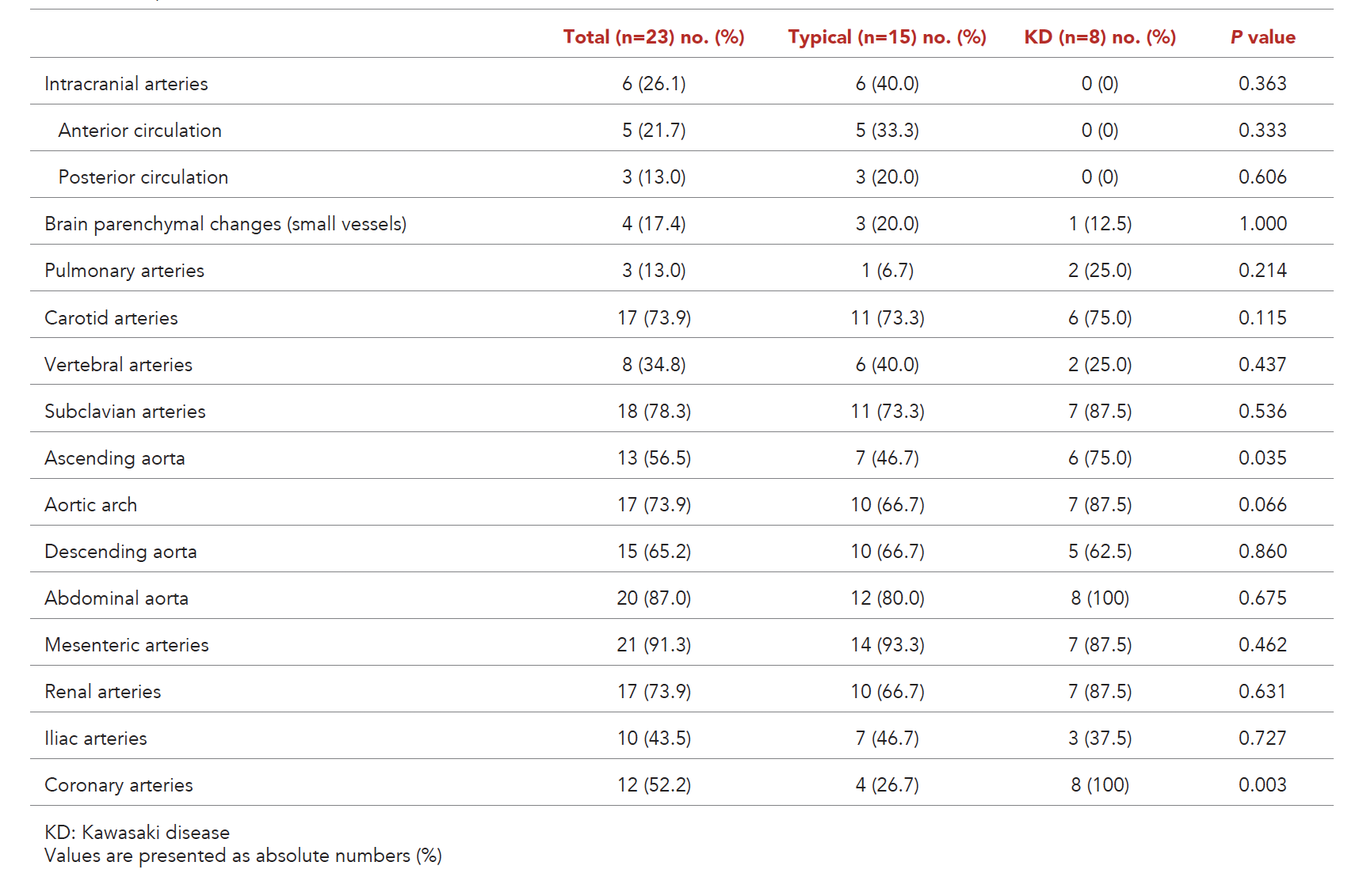
Twenty-one out of 23 patients (91%) had Type V angiographic patterns according to Hata’s classification (Supplementary Table S1). The remaining 2, who displayed Type I and Type IV, belonged to the typical c-TAK group. Overall, the most commonly involved vascular segments cumulatively were mesenteric arteries (21 patients, 91%), abdominal aorta (20 patients, 87%), subclavian arteries (18 patients, 78%), renal arteries (17 patients, 74%), aortic arch (17 patients, 74%), carotid arteries (17 patients, 74%), followed by descending (15 patients, 65%) and ascending aorta (13 patients, 57%) (Table 2). Not surprisingly, 100% (8 patients) of the c-TAK-KD group had CA involvement, compared to only 27% (4 patients) in the typical c-TAK group (P=0.003). None in the c-TAK-KD group had any intracranial artery involvement; only 1 of them had aneurysms in other medium-sized arteries (right brachial and left internal iliac arteries).
Table 2. Frequency of arterial territory involvement.
Treatment
The most commonly used immunosuppressive medications, besides glucocorticoids, were subcutaneous/oral methotrexate (MTX, 16 patients, 70%), followed by intravenous immunoglobulin (IVIg; 10 patients, 48%), intravenous cyclophosphamide (8 patients, 35%) and anti-tumour necrosis factor (aTNF, 8 patients, 35%) (Supplementary Table S2). All patients in the c-TAK-KD group received IVIg; 4 (50%) required MTX (after diagnosis of large-vessel vasculitis) and 3 (38%) required aTNF. Only 2 patients (9%) in the entire cohort received intravenous tocilizumab, and 1 of them (4%) received rituximab. Both of these patients were from the typical c-TAK group.
Outcomes
Sixteen patients (70%) in the entire cohort achieved CID with a median duration of 6 months (4–11) from treatment initiation (Table 3); a third of the cohort (8 patients, 35%) achieved CRM, with a median duration of 43 months (35–60) from treatment initiation. About a third of patients (8 patients, 35%) suffered at least 1 flare after achieving CID; the median duration from CID to flare was 13 months (4–17).
Table 3. Outcomes.
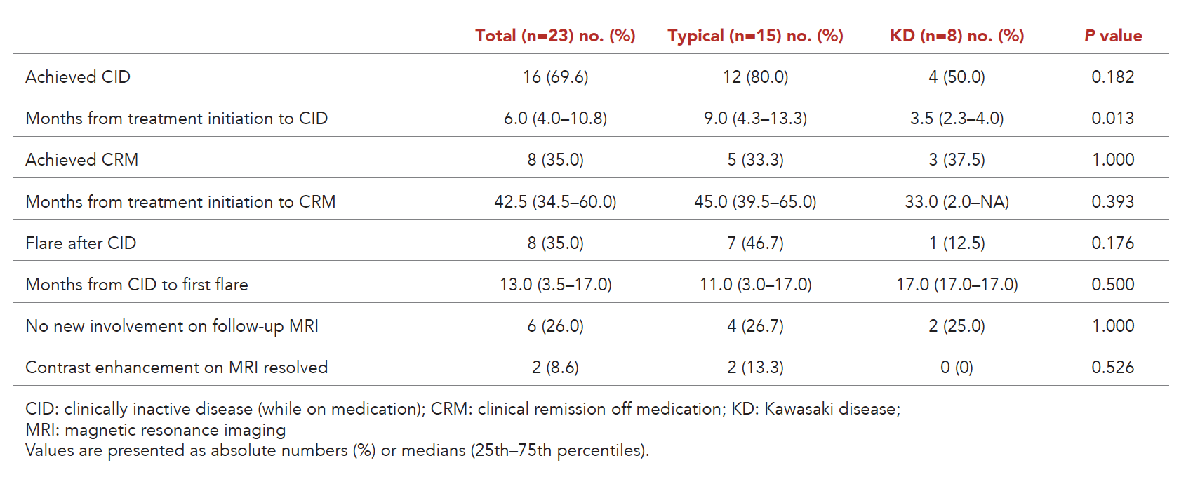
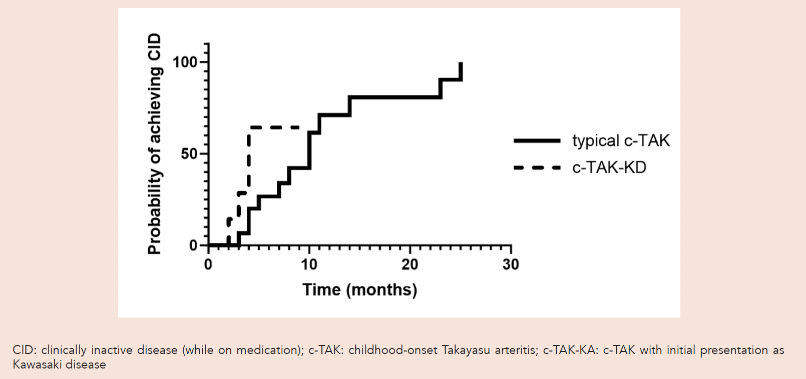
The typical c-TAK group had a higher percentage of patients achieving CID (80%) compared to the c-TAK-KD group (50%), although this did not reach statistical significance (P=0.182). The c-TAK-KD group took a significantly shorter duration to achieve CID than the typical c-TAK group (Fig. 1, Table 3). The proportion of patients who achieved CRM was similar between the 2 groups (5 patients [33%] in the typical c-TAK group vs 3 patients [38%] in the c-TAK-KD group, P=0.596). Among the patients who achieved CID, there was a higher percentage of patients who flared in the typical c-TAK group (7 patients, 47%) compared to the c-TAK-KD group (1 patient, 13%), although this did not reach statistical significance (P=0.176).
Fig. 1. Kaplan-Meyer curves of achieving inactive disease on medication (CID).
The radiological course of the disease did not seem to mirror the clinical course in our cohort completely. Out of all patients, only 6 patients (26%) showed no new area of involvement in the follow-up MRA (Table 3). Even fewer patients (2, 9%) achieved resolution of previously noted contrast enhancement in at least one follow-up MRA.
All 8 patients in the c-TAK-KD group developed coronary changes, among which 5 (62.5%) had medium- to giant aneurysms (Supplementary Table S3). In 4 (50%) of the 8 patients, the coronary changes eventually returned to normal on follow-up 2D echocardiogram, with a median duration of 5.5 (1.5–29.0) months. Among those with coronary aneurysms (5 patients), 2 (40%) patients had medium-sized aneurysms, and both achieved normalisation of their coronary arteries on follow-up. Three (60%) patients had giant aneurysms; 1 passed away from myocardial infarction, another had a persistent giant aneurysm at the end of follow-up, whereas the third had partial resolution with a moderate aneurysm at the end of follow-up. The remaining patients had only coronary dilatations, all of whom had normalisation of coronary arteries except for 1 who passed away from non-cardiac cause.
DISCUSSION
To our knowledge, our study is the first to analyse a c-TAK cohort in Southeast Asia with unique clinical characteristics. This study is also the first to describe 2 distinct groups of patient types: typical c-TAK and c-TAK-KD, that initially presented with KD features.
At first glance, our cohort seemingly deviated from previous studies regarding female preponderance (43.5% vs 64–79%),32,34-43 as well as the age of onset (median 3.8 years vs 9.2–16 years). This is in part due to skewing by the c-TAK-KD subgroup, who presented at a younger age and trended towards a male preponderance (Table 1); excluding that, the typical c-TAK cohort had sex distribution and age of onset closer to previous cohorts. Expectedly, all c-TAK-KD patients had an initial presentation of fever, and most had multiple KD features, which were otherwise absent in most typical c-TAK patients. They also had significantly higher inflammatory markers at presentation, as opposed to the typical c-TAK patients, who tended to present at a convalescent rather than an inflammatory stage. This group of patients was initially managed with IVIg for KD, but was refractory despite second-line treatment (either second dose of IVIg or pulse methylprednisolone, or both), requiring further immunosuppressants, and eventually found to have angiographic evidence of large-vessel vasculitis. It is unknown if this group of patients constitutes a distinct clinical entity, but all of them still fulfilled the EULAR/PRINTO/PReS and ACR/EULAR classification criteria for TAK.3,24 Kierzkowska et al. reported a 7-month-old female infant with an initial diagnosis of refractory KD who was later found to have abdominal aorta aneurysms.7 Lee et al. described a 4-year-old female with initial KD features and coronary aneurysms whose further imaging revealed diffuse aortitis.8 Shimura and colleagues also reported a 2-year-old boy with initial incomplete KD but had rapid deterioration, leading to death. Autopsy results indicated multiple sites of large-vessel stenosis, resulting in cerebral and renal infarctions.9 All 3 cases presented with an acute inflammatory state and fulfilled the criteria for KD diagnosis, but the diagnosis of c-TAK was only ascertained when the course of illness was atypical and further evaluations revealed other large vessel involvement. A recent report by Jin et al. described 10 patients with infantile-onset TAK (onset before 3 years old), more than half of whom were initially diagnosed and treated with KD, leading to significant morbidity and mortality.10 In patients with KD of an atypical course, a differential diagnosis of c-TAK should be considered, especially when patients are refractory to both first and second-line treatments.
Excluding the c-TAK-KD group, our typical c-TAK patients also differed from other cohorts in their clinical characteristics. Paediatric patients have been known to present with non-specific symptoms at a higher frequency than adults.2,34,43,44 However, our patients did not often present with constitutional symptoms like weight loss (3 patients, 20%) or malaise (2 patients, 13%). Comparatively, c-TAK cohorts in both the West and East Asia regions have significantly higher proportions of patients presenting with these constitutional symptoms (Fig. 2). Notably, our c-TAK patients also presented less often with hypertension (7 patients, 47%) compared to previously reported studies (57–100%).37,41 However, this did not preclude using the EULAR/PRINTO/PReS classification of c-TAK despite hypertension being one of the features listed. All our patients fulfilled both the EULAR/PRINTO/PReS and ACR/EULAR 2022 classification criteria.
None of our patients presented with claudication. In contrast, all cohorts in the current literature described patients presenting with claudication (Fig. 2). Apart from the UK cohort, all other cohorts, including West and East Asian cohorts, had more than 20% of the patients presenting with claudication.32,34-43 This is despite a relatively high proportion of patients with arterial involvement of the subclavian and iliac arteries (Table 2). On the other hand, our cohort of patients had a significantly higher proportion of patients presenting with stroke (33% vs 0–18%), all of whom had involvement of their intracranial arteries. This highlights c-TAK as an important differential diagnosis for ischaemic stroke in young patients with central nervous system (CNS) arteriopathies, in addition to infectious, cardiac, metabolic, haematologic and genetic causes.45 Evaluation of the aorta and its major branches should be considered in these cases.
Fig. 2. Comparison of clinical features across major published cohorts of childhood-onset Takayasu arteritis (excluding patients with initial presentation as Kawasaki disease).

In terms of angiographic patterns, the majority of our patients presented with Type V (21 patients, 91%), which is generally comparable to other cohort studies, although the latter did not have such overwhelming percentages (25–76%, Fig. 3).35,40 In addition, the most commonly involved territory in the entire cohort was the mesenteric arteries, followed by the abdominal aorta. Looking at differences among the subgroups, the c-TAK-KD patients had no intracranial involvement, and unsurprisingly, all had involvement of the CA. Interestingly, the c-TAK-KD group had a significantly greater involvement of the ascending aorta than the typical c-TAK group (6 patients [75%] vs 7 patients [47%], P=0.035).
Fig. 3. Comparison of angiographic classification types across major published cohorts of childhood-onset Takayasu arteritis.
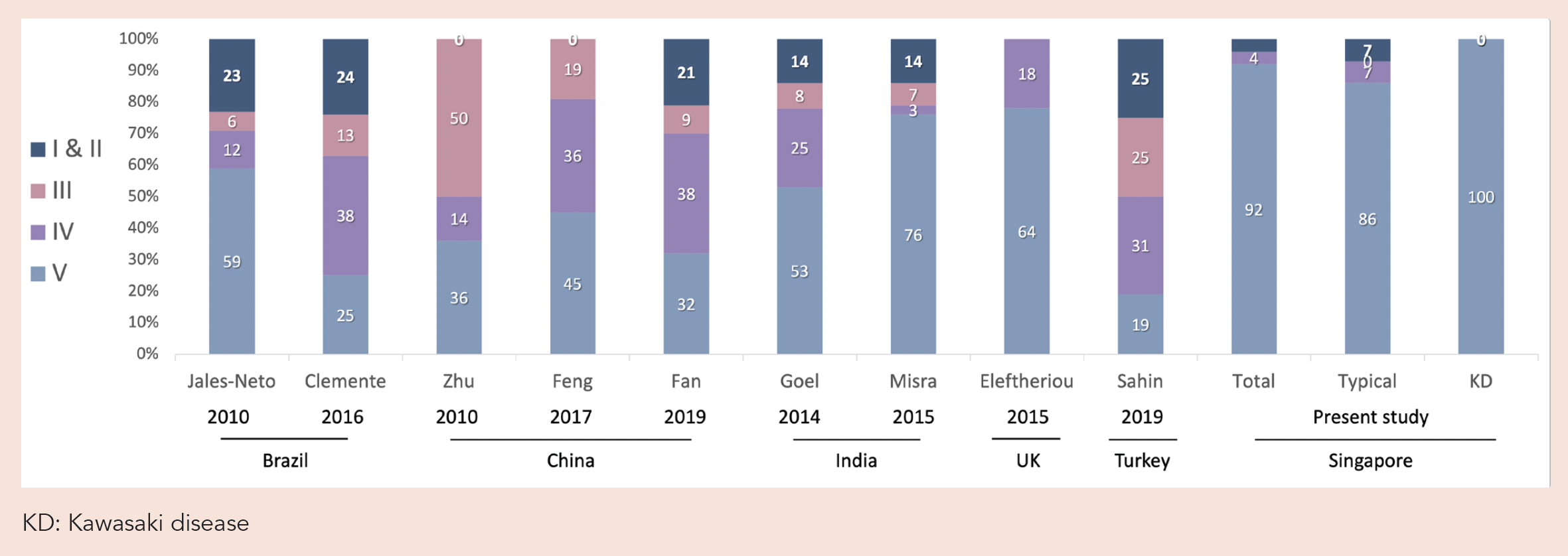
Even though most patients in our cohort were ethnically Chinese, the clinical characteristics were markedly different from various reports from China,10,16,17,23 from presenting symptoms to angiographic types (Figs. 1 and 2). This again echoes the epidemiological variability of c-TAK across populations and geographic regions, highlighting the uniqueness of our Southeast Asian cohort.
TAK is commonly seen in East Asia, where the prevalence of Mycobacterium tuberculosis (TB) infection is high, with various studies showing an association between TB infection and TAK.46,47 Half of our patients who had testing had a positive Mantoux test, but only 1 (7%) had positive IGRA testing. This discrepancy could be due to the nationwide Bacilli Calmette-Guerin vaccination at birth. In addition, autoantibodies and autoreactive T cells against human 60kDa heat shock protein (HSP) are involved in the pathogenesis of TAK, and these have been shown to cross-react with the mycobacterial 65kDa HSP, causing a false-positive Mantoux result.48 Nevertheless, none of our patients had active TB infection.
There is no recognised evidence-based recommendation for c-TAK treatment thus far. As such, there is a variation of preferred immunosuppressive agents for c-TAK in institutions worldwide. Treatment decisions for patients in our cohort were made at the discretion of the attending rheumatologists, although references were drawn from existing adult large-vessel vasculitis management guidelines by EULAR and ACR. Other than the obvious use of IVIg for c-TAK-KD, MTX was the most commonly used non-steroidal immunosuppressive agent in our cohort, which corresponds with the majority of previously published studies. This is compared to mycophenolate mofetil in an Indian study and azathioprine in a Turkish cohort.39,43 In our cohort, MTX was followed by cyclophosphamide in the typical c-TAK group (7 patients, 47%) and infliximab in the c-TAK-KD group (3 patients, 38%). Notably, the outcomes were not significantly different between the typical c-TAK and c-TAK-KD groups. Both groups had similar rates of achieving CRM during treatment, although the former trended towards a higher flare rate after inactive disease.
Previous studies of KD showed that close to 25% of patients developed CA lesions if not treated with IVIg.6 In comparison, patients in our c-TAK-KD cohort universally developed some degree of coronary lesions. Five (63%) of them received timely administration of the first dose of IVIg (Day 3 to 7 from fever onset); despite this, 4 developed medium- to giant coronary aneurysms (Supplementary Table S3). This observation, together with findings of large-vessel involvement, suggests that they are a clinically distinct group from the “run-of-the-mill” KD patients.
Lastly, although many of our patients achieved CID, the resolution of radiological features did not seem to correspond. Only 2 (9%) of our patients had a resolution of enhancement on MRA. However, the rest of the patients remained asymptomatic throughout their follow-up, suggesting a discrepancy between MRA findings and disease activity. In 1 meta-analysis, the reported utility of vessel wall thickening and enhancement by MRA to detect active disease in TAK varied across studies.49 Sartorelli et al. found that MRA had a 50% concordance with clinical assessment of disease activity and that vessel wall enhancement did not predict stenosis at follow-up in 2 years.50 Kato et al. compared 2 groups of TAK patients with active and inactive diseases, respectively, and showed that late gadolinium enhancement on MRA was useful in detecting the distribution of vessel wall involvement but was unable to accurately assess disease activity.51 An MRA scoring system with a combination of abnormal findings was proposed, but this was not validated in c-TAK cohorts.52 These findings emphasised the limitation of MRA imaging for disease monitoring in c-TAK and cautioned against the sole dependence on MRA to guide treatment.
This study is not without limitations. The sample size is small, but to our knowledge, this is the largest study on c-TAK to date in a Southeast Asian paediatric population. We acknowledge the potential for referral bias in this single-centre study; however, our centre—being the only children’s hospital in Singapore, with the biggest paediatric rheumatology clinic—helps to ensure its representativeness of the local population. Although we use a validated instrument (PVAS) to measure disease activity, other measurement tools exist.53,54 There is ultimately no universally accepted disease activity measure for childhood TAK yet. We had the availability of repeat MRA imaging in most patients, but FDG-PET scans were not routinely performed in our institution. Hence, we cannot compare and correlate findings between both imaging modalities. Lastly, we describe a distinct group of c-TAK-KD patients, but due to the small sample size, we could not match the patients with typical c-TAK for a more robust comparison between the 2 groups.
CONCLUSION
Our c-TAK cohort has unique characteristics compared to the Western and East Asian cohorts. It also highlights the heterogeneity in demographic, clinical, and angiographic characteristics of c-TAK in different geographic locations. The high proportion of CNS involvement and stroke in our cohort prompts c-TAK as an important differential diagnosis in young patients with stroke. Our observation of a distinct group of patients who initially presented with KD features has not previously been described. Early recognition of these entities and anticipating disparate treatment outcomes are essential to help reduce morbidity of our c-TAK patients through timely immunosuppression. Future studies would benefit from a longer follow-up duration and larger sample size to better understand this subgroup of c-TAK patients.
Supplementary material
Supplementary Table S1. Angiographic classification type.
Supplementary Table S2. Treatment.
Supplementary Table S3. Clinical characteristics and treatments of c-TAK-KD patients.
REFERENCES
- Aeschlimann FA, Yeung RS, Laxer RM. An update on childhood-onset Takayasu arteritis. Front Pediatr 2022;10:872313.
- Misra DP, Rathore U, Kopp CR, et al. Presentation and clinical course of pediatric-onset versus adult-onset Takayasu arteritis—a systematic review and meta-analysis. Clin Rheumatol 2022;41:3601-13.
- Ozen S, Pistorio A, Iusan S, et al. EULAR/PRINTO/PRES criteria for Henoch-Schonlein purpura, childhood polyarteritis nodosa, childhood Wegener granulomatosis and childhood Takayasu arteritis: Ankara 2008. Part II: Final classification criteria. Ann Rheum Dis 2010;69.
- Millan P, Gavcovich TB, Abitbol C. Childhood-onset Takayasu arteritis. Curr Opin Pediatr 2022;34:223-8.
- Vanoli M, Daina E, Salvarani C, et al. Takayasu’s arteritis: a study of 104 Italian patients. Arthritis Rheum 2005;53:100-7.
- McCrindle BW, Rowley AH, Newburger JW, et al. Diagnosis, treatment, and long-term management of Kawasaki disease: a scientific statement for health professionals from the American Heart Association. Circulation 2017;135:e927-99.
- Kierzkowska B, Lipińska J, Barańska D, et al. Takayasu’s arteritis mimicking Kawasaki disease in 7-month-old infant, successfully treated with glucocorticosteroids and intravenous immunoglobulins. Rheum Int 2012;32:3655-9.
- Lee M, Meidan E, Son M, et al. Coronary artery aneurysms in children is not always Kawasaki disease: a case report on Takayasu arteritis. BMC Rheumatol 2021;5:1-6.
- Shimura K, Fukushima R, Tanaka M. A case of Takayasu arteritis treated as incomplete Kawasaki disease 8 months ago. Pediatr Int 2023;65:e15586.
- Jin J, Zhao Y, Gao X, et al. A retrospective study of infantile-onset Takayasu arteritis: experience from a tertiary referral center in China. Front Cardiovasc Med 2024;11.
- Millan P, Gavcovich TB, Abitbol C. Childhood-onset Takayasu arteritis. Curr Opin Pediatr 2022;34:223-8.
- Aeschlimann FA, Raimondi F, Leiner T, et al. Overview of Imaging in Adult- and Childhood-onset Takayasu Arteritis. J Rheumatol 2022;49:346-57.
- Aeschlimann FA, Eng SWM, Sheikh S, et al. Childhood Takayasu arteritis: disease course and response to therapy. Arthritis Res Ther 2017;19:255.
- Clemente G, Hilário MO, Len C, et al. Brazilian multicenter study of 71 patients with juvenile-onset Takayasu’s arteritis: clinical and angiographic features. Rev Bras Reumatol Engl Ed 2016;56:145-51.
- Eleftheriou D, Varnier G, Dolezalova P, et al. Takayasu arteritis in childhood: retrospective experience from a tertiary referral centre in the United Kingdom. Arthritis Res Ther 2015;17:36.
- Fan L, Zhang H, Cai J, et al. Clinical course and prognostic factors of childhood Takayasu’s arteritis: over 15-year comprehensive analysis of 101 patients. Arthritis Res Ther 2019;21:31. Erratum in: Arthritis Res Ther 2019;21:55.
- Feng Y, Tang X, Liu M, et al. Clinical study of children with Takayasu arteritis: a retrospective study from a single center in China. Pediatr Rheumatol Online J 2017;15:29.
- Goel R, Kumar TS, Danda D, et al. Childhood-onset Takayasu arteritis — experience from a tertiary care center in South India. J Rheumatol 2014;41:1183-9.
- Jales-Neto LH, Levy-Neto M, Bonfa E, et al. Juvenile-onset Takayasu arteritis: peculiar vascular involvement and more refractory disease. Scand J Rheumatol 2010;39:506-10.
- Misra DP, Aggarwal A, Lawrence A, et al. Pediatric-onset Takayasu’s arteritis: clinical features and short-term outcome. Rheum Int 2015;35:1701-6.
- Sahin S, Hopurcuoglu D, Bektas S, et al. Childhood-onset Takayasu arteritis: A 15-year experience from a tertiary referral center. Int J Rheum Dis 2019;22:132-9.
- Szugye HS, Zeft AS, Spalding SJ. Takayasu Arteritis in the pediatric population: a contemporary United States-based single center cohort. Pediatr Rheumatol Online J 2014;12:21.
- Zhu WH, Shen LG, Neubauer H. Clinical characteristics, interdisciplinary treatment and follow-up of 14 children with Takayasu arteritis. World J Pediatr 2010;6:342-7.
- Grayson PC, Ponte C, Suppiah R, et al. 2022 American College of Rheumatology/EULAR classification criteria for Takayasu arteritis. Arthritis Rheumatol 2022;74:1872-80.
- Tan JHT, Fun HS, Arkachaisri T. Paediatrics rheumatology clinic population in Singapore: The KKH experience. Proceedings of Singapore Healthcare 2012;21:265-71.
- Clinical Testing Guidance for Tuberculosis: Tuberculin Skin Test. https://www.cdc.gov/tb/hcp/testing-diagnosis/tuberculin-skin-test.html. Accessed 18 July 2025.
- Hata A, Noda M, Moriwaki R, et al. Angiographic findings of Takayasu arteritis: new classification. Int J Cardiol 1996;54:S155-63.
- Dolezalova P, Price-Kuehne FE, Özen S, et al. Disease activity assessment in childhood vasculitis: development and preliminary validation of the Paediatric Vasculitis Activity Score (PVAS). Ann Rheum Dis 2012;72:1628-33.
- Wang J, Dai X, Ma L, et al. Efficacy and safety of tofacitinib versus leflunomide with glucocorticoids treatment in Takayasu arteritis: A prospective study. Semin Arthritis Rheum 2022;55:152018.
- Tian Y, Chen Y. Stroke in Takayasu arteritis with concomitant tuberculosis: an unusual pediatric case report. BMC Pediatr 2022;22:50.
- Kong X, Sun Y, Dai X, et al. Treatment efficacy and safety of tofacitinib versus methotrexate in Takayasu arteritis: a prospective observational study. Ann Rheum Dis 2022;81:117-23.
- Aeschlimann FA, Eng SW, Sheikh S, et al. Childhood Takayasu arteritis: disease course and response to therapy. Arthritis Res Ther 2017;19:255.
- McCrindle BW, Rowley AH, Newburger JW, et al. Diagnosis, Treatment, and Long-Term Management of Kawasaki Disease: A Scientific Statement for Health Professionals From the American Heart Association. Circulation 2017;135:e927-99.
- Jales-Neto L, Levy-Neto M, Bonfa E, et al. Juvenile-onset Takayasu arteritis: peculiar vascular involvement and more refractory disease. Scand J Rheumatol 2010;39:506-10.
- Clemente G, Hilário MO, Len C, et al. Brazilian multicenter study of 71 patients with juvenile-onset Takayasu’s arteritis: clinical and angiographic features. Rev Bras Reumatol Engl Ed 2016;56:145-51.
- Zhu W-H, Shen L-G, Neubauer H. Clinical characteristics, interdisciplinary treatment and follow-up of 14 children with Takayasu arteritis. World J Pediatr 2010;6:342-47.
- Feng Y, Tang X, Liu M, et al. Clinical study of children with Takayasu arteritis: a retrospective study from a single center in China. Pediatr Rheumatol Online J 2017;15:29.
- Fan L, Zhang H, Cai J, et al. Clinical course and prognostic factors of childhood Takayasu’s arteritis: over 15-year comprehensive analysis of 101 patients. Arthritis Res Ther 2019;21:31. Erratum in: Arthritis Res Ther 2019;21:55.
- Goel R, Kumar TS, Danda D, et al. Childhood-onset Takayasu arteritis—experience from a tertiary care center in South India. J Rheumatol 2014;41:1183-9.
- Misra DP, Aggarwal A, Lawrence A, et al. Pediatric-onset Takayasu’s arteritis: clinical features and short-term outcome. Rheumatol Int 2015;35:1701-6.
- Szugye HS, Zeft AS, Spalding SJ. Takayasu arteritis in the pediatric population: a contemporary United States-based single center cohort. Pediatr Rheumatol Online J 2014;12:21.
- Eleftheriou D, Varnier G, Dolezalova P, et al. Takayasu arteritis in childhood: retrospective experience from a tertiary referral centre in the United Kingdom. Arthritis Res Ther 2015;17:36.
- Sahin S, Hopurcuoglu D, Bektas S, et al. Childhood‐onset Takayasu arteritis: A 15‐year experience from a tertiary referral center. Int J Rheum Dis 2019;22:132-9.
- Brunner J, Feldman BM, Tyrrell PN, et al. Takayasu arteritis in children and adolescents. Rheumatology (Oxford) 2010;49:1806-14.
- Janas AM, Barry M, Lee S. Epidemiology, causes, and morbidities of stroke in the young. Curr Opin Pediatr 2023;35:641-7.
- Magar MT, Kafle S, Poudel A, et al. Takayasu’s Arteritis and Its Association With Mycobacterium Tuberculosis: A Systematic Review. Cureus 2021;13:e16927.
- Pedreira ALS, Santiago MB. Association between Takayasu arteritis and latent or active Mycobacterium tuberculosis infection: a systematic review. Clin Rheumatol 2020;39:1019-26.
- Castillo-Martínez D, Amezcua-Castillo LM, Granados J, et al. Is Takayasu arteritis the result of a Mycobacterium tuberculosis infection? The use of TNF inhibitors may be the proof-of-concept to demonstrate that this association is epiphenomenal. Clin Rheumatol 2020;39:2003-9.
- Barra L, Kanji T, Malette J, et al. Imaging modalities for the diagnosis and disease activity assessment of Takayasu’s arteritis: a systematic review and meta-analysis. Autoimmun Rev 2018;17:175-87.
- Sartorelli S, Di Chio M, Baldissera E, et al. SAT0292 Clinical, imaging and laboratory variables fail to predict morphological vascular progression in takayasu arteritis. Ann Rheum Dis 2014;73:698-9.
- Kato Y, Terashima M, Ohigashi H, et al. Vessel Wall Inflammation of Takayasu Arteritis Detected by Contrast-Enhanced Magnetic Resonance Imaging: Association with Disease Distribution and Activity. PLoS One 2015;10:e0145855.
- Jiang L, Li D, Yan F, et al. Evaluation of Takayasu arteritis activity by delayed contrast-enhanced magnetic resonance imaging. Int J Cardiol 2012;155:262-7.
- Aydin SZ, Yilmaz N, Akar S, et al. Assessment of disease activity and progression in Takayasu’s arteritis with Disease Extent Index-Takayasu. Rheumatology (Oxford) 2010;49:1889-93.
- Misra R, Danda D, Rajappa SM, et al. Development and initial validation of the Indian Takayasu clinical activity score (ITAS2010). Rheumatology (Oxford) 2013;52:1795-801.
The SingHealth Centralised Institutional Review Board (2019/2961 and 2019/2194) approved this study and waived the need for informed consent due to the nature of retrospective data review from the registry.
The authors declare there are no affiliations with or involvement in any organisation or entity with any financial interest in the subject matter or materials discussed in this manuscript.
Professor Thaschawee Arkachaisri, Rheumatology and Immunology Service, Department of Pediatric Subspecialties, KK Women’s and Children’s Hospital, 100 Bukit Timah Road, Children’s Tower, Singapore 229899. Email: [email protected]
