

ABSTRACT
Haemophagocytic lymphohistiocytosis (HLH) is a rare, life-threatening hyperinflammatory state characterised by inappropriate survival of immune cells, cytokine storm, profound cytopenias, and rapidly progressive multi-organ failure. In adults with secondary HLH, the most common triggers include haematological malignancy, autoimmune/autoinflammatory conditions, and infections. In Asian contexts, both malignancy-induced (with higher rates of NK/T-cell lymphoma than B-cell lymphoma) and Epstein-Barr virus-induced HLH are particularly common. Additionally, there is a distinct “Asian” variant of intravascular B-cell lymphoma with no cutaneous manifestations that tends to manifest with HLH and may require random skin biopsies to diagnose. Diagnosis can be supported by the HLH-2004 diagnostic and therapeutic guidelines for haemophagocytic lymphohistiocytosis or the H-score in adult HLH, and subsequent management involves high-dose corticosteroids with targeted immunomodulatory treatments or chemotherapy directed at the underlying condition. In this narrative review article, the authors provide a comprehensive overview of HLH epidemiology, diagnosis, and management strategies with a practical approach for frontline clinicians practising in Asian contexts.
CLINICAL IMPACT
What is New
- This review provides a practical clinical approach to patients with suspected haemophagocytic lymphohistiocytosis (HLH) based on clinical assessments, laboratory and bone marrow findings.
- Evidence-based management options based on aetiological classification of HLH are discussed.
Clinical Implications
- Management options for HLH include high-dose corticosteroids, intravenous immunoglobulin, interleukin-1 receptor antagonists, and etoposide.
Vignette:
A 55-year-old Chinese lady was admitted for pyrexia of unknown origin for 3 weeks, associated with a 5 kg weight loss. She had no rashes and no infective, autoimmune, or other systemic symptoms. On admission, she had fluid-refractory hypotension requiring inotropic support in the intensive care unit. Her physical examination was otherwise unremarkable. Laboratory investigations revealed severe pancytopenia (white blood cell 0.9 x 10⁹/L, haemoglobin 6.5 g/dL, platelet 16 x 109/L), transaminitis (aspartate transaminase 500 U/L, alanine transaminase 720 U/L, alkaline phosphatase 875 IU/L), hyperferritinaemia (ferritin 55,000 ng/mL), hypertriglyceridaemia (4.1 mmol/L) and hypofibrinogenaemia (1.1 g/L). The coagulation panel was normal. The peripheral blood film did not show early cells or features of haemolysis. A whole-body positron-emission tomography-computed tomography scan did not reveal suspicious masses or significant lymphadenopathy. Bone marrow trephine biopsy revealed haemophagocytosis, with no evidence of haematological malignancy. The patient was diagnosed with idiopathic haemophagocytic lymphohistiocytosis (HLH) and treated with intravenous pulsed methylprednisolone. However, in the following days, the patient deteriorated, requiring dual inotropic support and intubation for mechanical ventilation. The deterioration was attributed to a systemic hyperinflammatory state, with possible nosocomial pneumonia. Despite supportive and broad-spectrum antimicrobial treatment, she eventually succumbed to her illness. A post-mortem examination was performed, revealing that the patient had intravascular B-cell lymphoma. The case was subsequently presented at a mortality and morbidity meeting, and the team reviewed the literature, which showed an elevated incidence of malignancy-associated HLH in Asian populations (with NK/T-cell more than B-cell lymphoma). Notably, the haemophagocytic subtype of intravascular large B-cell lymphoma, historically termed the “Asian variant”, is particularly relevant in Asian populations; it may present without cutaneous involvement and tends to manifest with HLH, which was consistent with the patient’s case. In such cases, random skin biopsies could have been helpful for diagnosis. The team also reflected that continued aggressive pursuit of the rare causes of HLH remains necessary even if the standard infective/autoimmune/malignancy workup is unyielding, while being mindful of the potential for immunosuppressive treatment to mask the underlying disease process.
INTRODUCTION
Haemophagocytic lymphohistiocytosis (HLH) is a life-threatening hyperinflammatory syndrome characterised by inappropriate survival and proliferation of cytotoxic T-cells and macrophages/histiocytes, leading to cytokine storm (uncontrolled systemic inflammatory response), haemophagocytosis, and end-organ damage.1 With the rare exception of familial HLH that is typically seen in the paediatric population, adult patients usually develop HLH in response to underlying infective triggers, occult or known malignancies, or autoimmune conditions. Macrophage activation syndrome (MAS) is considered a subset of secondary HLH that is triggered by underlying autoimmune conditions such as systemic juvenile idiopathic arthritis (sJIA), adult-onset Still’s disease (AOSD), or systemic lupus erythematosus (SLE).
Patients with HLH often present clinically with haemodynamic instability, fevers, and even multi-organ failure requiring intensive care. Laboratory investigations may reveal characteristic features of severe hyperferritinaemia, hypofibrinogenaemia, fasting hypertriglyceridaemia, and a hepatocellular pattern of transaminitis. These manifestations of HLH can mimic those of other systemic inflammatory states such as sepsis and systemic inflammatory response syndrome, resulting in HLH being frequently misdiagnosed or diagnosed late. This delay can lead to severe consequences, as timely treatment is crucial to improving patient outcomes.2 The use of criteria that rely on laboratory tests, many of which have significant overlap with other diseases, poses additional challenges.3
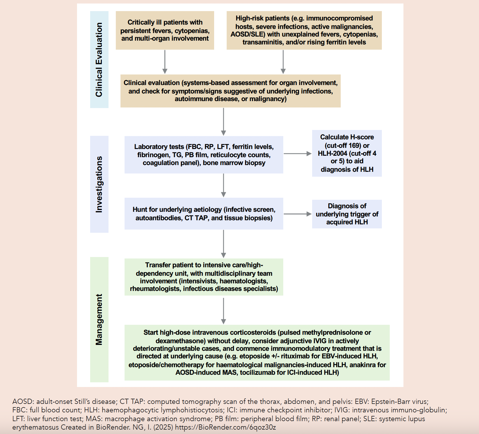
In this narrative review article, the authors focus on a practical approach for the diagnosis and management of HLH in the adult population for frontline clinicians (Fig. 1).
Fig. 1. Diagnostic and management algorithm for suspected cases of HLH.
METHODS
For the purposes of this narrative review article, the authors performed an updated PubMed search for articles from January 2000 to December 2025 using the following terms in title and abstract searches: “haemophagocytic lymphohistiocytosis” or “hemophagocytic lymphohistiocytosis” or “macrophage activation syndrome” and “adult”, and included only articles with full-text availability and article types that were original research, guidelines, or review articles. This yielded 296 articles, which were reviewed alongside the authors’ personal libraries of relevant articles on this topic.
RESULTS
Aetiology and disease pathophysiology
From molecular studies, the current postulation of HLH pathophysiology involves a probable underlying genetic predisposition to cytolytic pathway dysfunction or disruption, such that immune cell apoptosis (immune downregulation) is impaired, culminating in a vicious cycle of CD8+ T-lymphocyte and macrophage activation with cytokine release an overall sustained systemic hyperinflammatory state.1,4
HLH can occur in both the paediatric (<18 years) and adult populations, and can be primary or secondary. Primary HLH typically manifests in early childhood due to mutations in genes such as PRF1, UNC13D, STX11, and STXBP2 that disrupt the interactions among natural killer (NK) cells, CD8+ cytotoxic T-cells, and antigen-presenting cells.1 Consequently, these dysfunctional and deregulated cells produce increased levels of proinflammatory cytokines, causing systemic activation of macrophages and a systemic inflammatory state.5 Primary HLH can be further classified based on the inheritability of the genetic mutations involved (X-linked, recessive, or dominant), or the clinical syndrome it occurs in, such as Griscelli syndrome.5 Evidence has emerged of heterozygous mutations causing HLH as well. While most primary forms of HLH require 2 mutated copies (homozygous), a single heterozygous mutation in genes like RAB27A can increase the risk of developing secondary HLH or trigger a less common presentation of primary HLH, especially when combined with other mutations or factors.6,7 In the absence of a characterised genetic syndrome, such cases of primary HLH are commonly termed familial HLH.
Unlike its paediatric counterpart, adult-onset HLH is secondary to an acquired pathology. From epidemiological studies across the world (US, Europe, and Asia) of secondary HLH cases in adults, the 3 major causes of HLH are haematological malignancies (29–73%), infections (18–38%), and autoimmune/autoinflammatory conditions (0–21%) (Table 1).8–15 In Ramos-Casals et al.’s 2014 review of 2197 reported cases of HLH worldwide, it appears that Asian settings (China, South Korea, Japan, and Taiwan) had higher rates of malignancy-triggered HLH than the West (US and European countries), at approximately 50% versus (vs) 33%.16 In 2 cohort studies in China, malignancy-induced HLH was the predominant cause (42–46%), and T/NK-cell non-Hodgkin lymphoma outnumbered B-cell lymphoma.14,15 On the other hand, the proportion of autoimmune-related HLH was similar in both Asian and Caucasian cohorts.16 Infection remains a common trigger in most developed countries, with Epstein-Barr virus (EBV) being the predominant causative virus in Asia and the US, whereas human immunodeficiency virus (HIV) is more commonly the infective trigger in European countries.16 In 2 cohort studies in China, EBV infection was the most commonly identified infective trigger.14,15 Besides geographical location, there is also heterogeneity in the distribution of adult HLH aetiologies depending on the clinical setting (e.g. acute and critical care, specialist centres).
Table 1. Aetiologies of secondary HLH in adults.
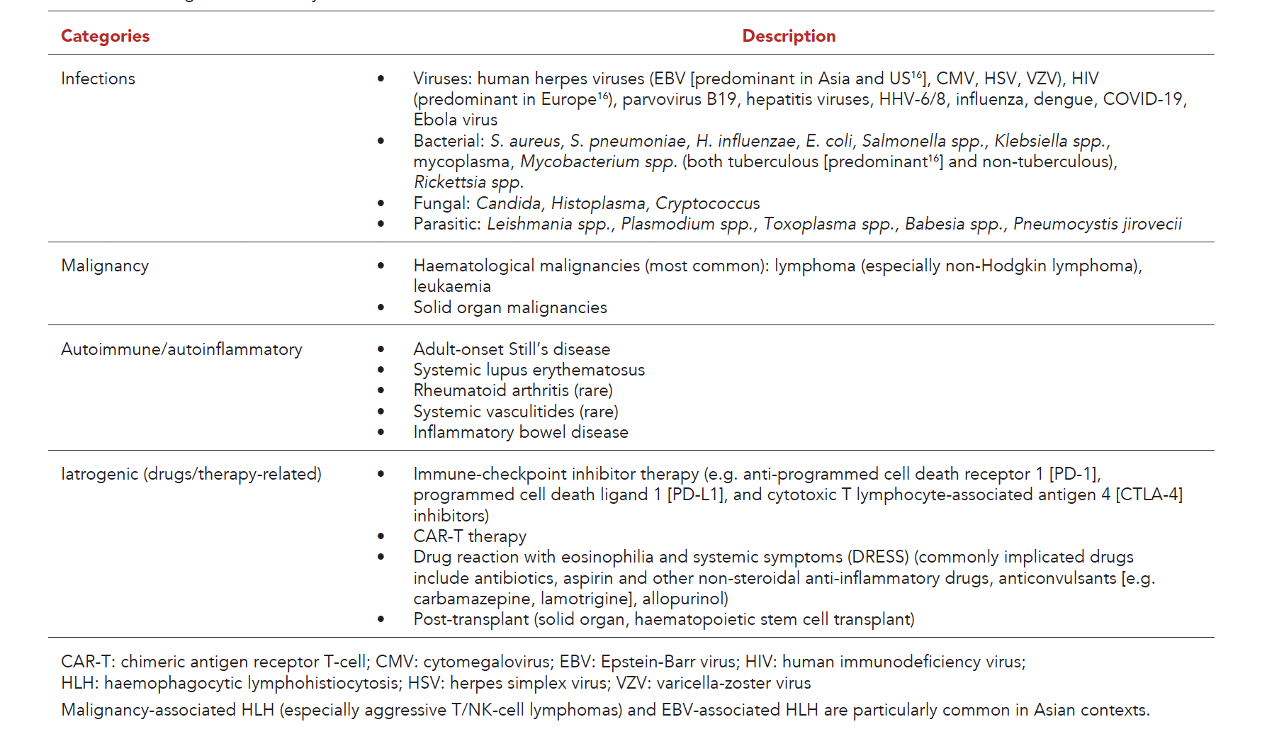
Iatrogenic causes of HLH are rare but increasingly recognised, ranging from post-transplant (solid organ or haematopoietic stem cell transplant) to drug reaction with eosinophilia and systemic symptoms (DRESS) and, most recently, chimeric antigen receptor T-cell (CAR-T)/bispecific T-cell engager (BiTE)/immune checkpoint inhibitor (ICI)-associated HLH.4,16-19 In the absence of known primary HLH-associated genetic mutations that can, by themselves, lead to HLH manifestations, an acquired “trigger” (infection, malignancy, autoimmune disease, and/or drugs) is often required to set off this hyperinflammatory state.1,4 Additionally, it is not uncommon to have patients with no identifiable cause of secondary HLH, with nearly 1 in 5 cases labelled as “idiopathic”.20
In adult HLH, a third of cases have more than 1 identified aetiology, for example, haematological malignancy or autoimmune disease with superimposed infection.16 Brisse’s threshold model best illustrates the current understanding of adult secondary HLH, which posits that endogenous components (genetic predisposition) and exogenous components (e.g. immunocompromised state, infective triggers, malignancy) are required to collectively tip the patient over a “threshold” beyond which fulminant HLH occurs.1,17
Clinical approach to identifying and diagnosing HLH
Clinicians should have a low threshold to evaluate for HLH in patients with pyrexia of unknown origin, as well as critically ill patients with fever, cytopenias involving 2 or more cell lines, and evidence of multi-organ involvement. Patients with HLH may present insidiously4; therefore, close monitoring for evolving HLH is warranted in high-risk patients, such as those with a known immunosuppressed state, severe infections, active malignancies, or associated autoimmune conditions (AOSD or SLE), who develop unexplained fevers, cytopenias, transaminitis, or rising ferritin levels, even if they appear clinically “well” or stable.
Patients with HLH can present with a myriad of multisystemic clinical manifestations related to the hyperinflammatory state/sequelae and/or underlying condition. These may include persistent unremitting fevers; haemodynamic instability (e.g. due to third-spacing from vasodilatory state or hypoalbuminaemia); neurological symptoms (e.g. confusion, ataxia, seizures, headaches, irritability); non-specific gastrointestinal symptoms (e.g. nausea/vomiting, abdominal pain, diarrhoea); hepatosplenomegaly; lymphadenopathy; as well as evidence of underlying cytopenias (manifesting as symptoms/signs of anaemia, bleeding diatheses from thrombocytopenia); liver dysfunction (manifesting as jaundice, altered mentation due to hepatic encephalopathy, and coagulopathy); renal dysfunction (with acute kidney injury potentially requiring renal replacement therapy); and/or acute respiratory distress syndrome (manifesting as respiratory failure).4,17
In patients with clinical manifestations suggestive of HLH, it is important to evaluate for clinical clues that may suggest underlying infections, rheumatic conditions, malignancy, and iatrogenic causes. For instance, clinicians should elicit infectious symptoms/signs such as respiratory symptoms (cough, rhinorrhoea, sore throat, breathlessness or pleuritic chest pain); gastrointestinal symptoms (nausea, vomiting, abdominal pain, diarrhoea); urinary tract symptoms (dysuria, urinary frequency and urgency, foul-smelling urine, haematuria); and skin rashes. Predisposing history and risk factors should also be elicited, including the presence of a known immunocompromised state (e.g. history of human immunodeficiency virus infection, or use of immunosuppressants); positive sick contacts; travel history; and high-risk sexual behaviours and exposures. In addition, to evaluate for associated autoimmune conditions, clinicians should assess for quotidian fevers, salmon-pink evanescent rashes and/or arthralgias/arthritis that wax and wane with fever, sore throat, clinical evidence of serositis (pleuritic chest pains, pleural effusions), hepatosplenomegaly, and/or lymphadenopathy, which may be seen in AOSD, and evaluate for symptoms/signs of connective tissue disease including inflammatory arthritis, alopecia, eye pain/redness, oral ulcers, malar rash or photosensitivity, sicca symptoms, Raynaud’s phenomenon, vasculitic rashes or cutaneous manifestations of SLE. Likewise, assess for B-symptoms (fever, night sweats, significant loss of appetite/weight) and generalised lymphadenopathy, which may be seen in lymphomas. It is equally important to review patients’ medication records to look for potential drug triggers associated with DRESS (these patients may also have eosinophilia and rashes). It is crucial to perform a review of drug history, and for patients with known cancers, the recent treatment history of immune checkpoint inhibitors or CAR-T therapies.
Table 2. Key investigations in suspected cases of secondary HLH in adults.
In all suspected cases of HLH, laboratory tests including full blood count, liver and renal function tests, serum ferritin levels, fibrinogen, fasting triglyceride (TG), peripheral blood film and reticulocyte counts, haemolytic screen (haptoglobin, lactate dehydrogenase [LDH], and indirect bilirubin), coagulation panel, and/or specialised biomarkers (soluble CD25, CD163, NK cell activity, cytokine panel) should be performed (Table 2).24 Although measurement of fasting triglyceride levels is suggested in conventional HLH criteria,21 it may not be practical in acutely ill patients. Non-fasting lipids are increasingly accepted in guidelines on routine cholesterol screening and cardiovascular risk prediction25; however, the accuracy of non-fasting lipids in HLH criteria remains to be established. As with other biochemical markers, TG should not be interpreted in isolation and is neither specific nor sensitive on its own to predict HLH. For instance, a recent study found that hypertriglyceridaemia was a common metabolic sequela that persisted in 1 in 5 intensive care unit patients even 3 months after their discharge.26 Although immune biomarkers such as those assessing T/NK-cell activity have increasingly been described, their use in routine clinical practice remains limited by cost considerations and lack of widespread test availability.24 Serum ferritin is an excellent screening biomarker: levels <500 µg/L have a 94% sensitivity in ruling out HLH,22 and very high levels (>10,000 µg/L) are highly suggestive of the diagnosis in paediatric HLH.23 However, extreme elevation of ferritin levels has lower diagnostic specificity in adults, in whom sepsis, liver failure, adult-onset Still’s disease and malignancy can also produce extreme hyperferritinaemia. Of note, C-reactive protein is typically elevated but lacks diagnostic specificity and can be blunted when interleukin (IL)-6 inhibitor therapy is used, whereas erythrocyte sedimentation rate (ESR) can be spuriously normal due to hypofibrinogenaemia in HLH.4,20
Bone marrow aspirate and trephine biopsy were traditionally considered to be important in the diagnostic evaluation of suspected HLH, in part due to their stated role in the original HLH diagnostic criteria. The utility of bone marrow evaluation is primarily to look for histopathological evidence of haemophagocytosis. However, bone marrow haemophagocytosis has poor sensitivity and specificity: it is absent in up to 40% of HLH patients because it appears late, and it can also occur secondary to infection, autoimmune disease, marrow failure, and transfusion.4,21,27
Table 3. Key HLH diagnostic criteria.
To facilitate the diagnosis of HLH, 3 sets of main criteria can be used: the HLH-2004 criteria22 (originally developed for diagnosis of primary/familial HLH, revised in 20241,28), H-score8 (developed for use in adult patients with secondary HLH), and European Alliance of Associations for Rheumatology/American College of Rheumatology/Paediatric Rheumatology International Trials Organisation 2016 MAS classification criteria29 (originally developed for use in MAS secondary to sJIA, and subsequently validated in adult patients with AOSD30) (Table 3). While the HLH-200422 and HLH-202428 criteria are often extrapolated for use in adult HLH, it must be recognised that their reportedly high sensitivity/specificity were derived from their performance in the original derivation cohort of paediatric familial HLH, and when the HLH-2004 criteria were applied to adult validation cohorts, their diagnostic accuracy was lower.31,32
Recently, Lachmann et al. performed a multicentre validation study of the HLH-2004 criteria, HLH-2024 criteria, and the H-score across 13 international secondary-HLH cohorts comprising both adult and paediatric patients. They found that the best-performing diagnostic criteria were the original HLH-2004 criteria (using decreased cut-off ≥4) (mean sensitivity 87%, mean specificity 86%), followed by HLH-2024 criteria (using decreased cut-off ≥4) (mean sensitivity 84%, mean specificity 88%), and then H-score (using cut-off ≥169) (mean sensitivity 82%, mean specificity 88%).25 On subgroup analysis of only intensive care unit patients, all 3 models demonstrated good diagnostic performance.25 For screening purposes, the authors found that a ferritin cut-off of 500 µg/L had 94% mean sensitivity in all cohorts to rule out secondary HLH.25 Bilston et al.’s 2022 multicentre study of adult HLH patients found that the HLH-2004 criteria (using cut-off ≥5) were more specific (93% vs 71%), but less sensitive (91% versus [vs] 96%) than H-score (using cut-off ≥169).31 In general, HLH-2004 performed better than the H-score for most aetiologies, with the exception of autoimmune and inflammatory aetiologies, for which the H-score had better sensitivity.31 In post-haematopoietic stem cell transplant recipients, a higher optimal H-score cut-off ≥233 was reported, due to a higher probability of presenting with post-transplant manifestations of fever, cytopenias, splenomegaly and systemic inflammatory response syndrome, which overlap with HLH criteria.31
In recent years, there have also been various criteria developed to assess for immune effector cell-associated haemophagocytic lymphohistiocytosis-like syndrome (IEC-HS), a severe form of haemotoxicity post-CAR T-cell therapy, including the MD Anderson 2018 criteria,33 the European Society for Blood and Marrow Transplantation (EBMT)/Joint Accreditation Committee of the International Society for Cell and Gene Therapy (ISCT) and EBMT/European Hematology Association 2022 best practice recommendations,34 and the IEC-HS 2023 criteria.35
In adults with undifferentiated HLH, the H-score offers a convenient, calculator-supported estimate of probability for clinicians, but it should be interpreted alongside the HLH-2004/HLH-2024 criteria; of note, its specificity is lower and diagnostic performance varies based on the cohort profile and underlying aetiology, so no single tool replaces clinical judgement and an active search for the trigger. The H-score (conventional cut-off ≥169) comprises important constituent items including underlying immunosuppressed state, fever, presence of hepatosplenomegaly, cytopenias, hyperferritinaemia, hypertriglyceridaemia, hypofibrinogenaemia, and elevated aspartate transaminase levels, as well as bone marrow evidence of haemophagocytosis.8
The extent and sequence of aetiological workup should be based on clinical suspicion of underlying aetiology, patients’ preferences and goals of care, and resource availability or cost considerations. In general, an infective workup could include serologies for EBV, herpes simplex virus, varicella-zoster virus, and human immunodeficiency virus, as well as a dengue screen, COVID-19/respiratory viral swabs, mycoplasma serology, and other specific microbiological or serological tests for bacterial, fungal, and/or parasitic infections, depending on the index of clinical suspicion. Antinuclear antibodies, anti-double-stranded DNA, complement C3/C4, and/or rheumatoid factor/anti-cyclic citrullinated peptide, or antineutrophil cytoplasmic antibodies could be evaluated for the possibility of connective tissue diseases. A computed tomography (CT) scan of the neck, thorax, abdomen, and pelvis, or even a positron-emission tomography-CT scan, could be performed to look for evidence of occult malignancies, organomegaly, lymphadenopathy, infected collections, or even vessel wall abnormalities, which may suggest intravascular lymphoma or vasculitis. Bone marrow evaluation is also helpful to look for signs of haematological malignancies (lymphoma, leukaemia). Besides bone marrow evaluation, biopsy of affected/abnormal tissues (identified clinically or on imaging modalities) may help with the diagnosis of the underlying condition,4 such as excisional lymph node biopsy to evaluate for underlying lymphoma, or bronchoalveolar lavage/transbronchial lung biopsy or liver biopsy to exclude infections and malignancies. In particular, there is a highly fatal yet highly elusive form of lymphoma known as intravascular large B-cell lymphoma, which can present with a haemophagocytic variant in Asian populations characterised by fevers, hepatosplenomegaly, thrombocytopenia, and bone marrow involvement with non-neoplastic haemophagocytic histiocytes.36 If left undiagnosed or untreated, it is highly aggressive with a median survival time of 2–8 months.36 Although this subtype of intravascular lymphoma often has no obvious cutaneous involvement, limited evidence has shown that random skin biopsies (RSBs) may be helpful to diagnose this insidious entity, especially in Asian patients with a high-risk phenotype (pyrexia of unknown origin, diagnosed HLH, thrombocytopenia, and markedly elevated LDH).37 RSBs (either incisional or telescopic punch biopsies) should be performed over 3 or 4 different anatomical sites, usually over the thigh, posterior arm, and abdominal wall, as well as cherry angiomas if they are present.38 While life-saving immunosuppressive treatment should not be unduly delayed, when possible, it is preferable for PET-CT scans and tissue biopsies to be performed prior to initiation of high-dose glucocorticoids, which have the potential to reduce diagnostic yield, especially in the context of steroid-sensitive lymphomas.
Management strategies for HLH
Management of patients with HLH involves supportive care in a high-dependency or intensive care unit, with multidisciplinary team involvement usually comprising intensivists, haematologists, rheumatologists, and infectious disease specialists.
Table 4. Common treatment options for HLH.
Initial immunomodulatory treatment for all HLH cases (Table 4) typically involves high-dose glucocorticoids in the form of either intravenous “pulsed” methylprednisolone (500 mg–1 g/day for 3–5 days) followed by maintenance prednisolone (1 mg/kg/day) on a tapering regimen, or a dexamethasone regimen based on the HLH-94 regimen (10 mg/m2 for 2 weeks, 5 mg/m2 for 2 weeks, 2.5 mg/m2 for 2 weeks, 1.25 mg/m2 for 1 week, then taper off by week 8).39 Of note, dexamethasone may be the preferred option in patients with central nervous system (CNS) involvement due to better CNS penetration.27 Intravenous immunoglobulin (1 g/kg/day for 2 days, which can be repeated after 2 weeks) may be considered as an adjunctive “bridging” treatment option for deteriorating or unstable patients.19,21
The choice of treatment depends on the suspected underlying trigger. Infection-associated HLH requires prompt initiation of antimicrobial treatment. Specifically, in patients with EBV-associated HLH, rituximab can reduce the EBV-infected B-cell reservoir and is a useful adjunct, but etoposide-based therapy remains the backbone for high-risk or refractory disease, especially as EBV typically resides in T/NK compartments.40,41 For patients with MAS secondary to AOSD, IL-1 receptor antagonist (anakinra), calcineurin inhibitor (cyclosporine), and/or interferon (IFN)-γ inhibitor (emapalumab) should be considered.42 For MAS secondary to other rheumatic diseases, guideline-directed therapeutic options for severe/life-threatening manifestations can be considered, for instance, cyclophosphamide or rituximab, on top of hydroxychloroquine in SLE.43 Refractory MAS may be treated with etoposide.19 For patients with malignancy, definitive chemotherapeutic or other forms of treatment should be commenced as soon as the patient is stabilised, as malignancy-associated HLH will continue to recur if the underlying malignancy remains active. Nonetheless, the exact timing at which cancer treatment (which is also highly immunosuppressive) should be commenced depends on the clinician’s experience and patient contextual factors. In general, the prognosis of malignancy-associated HLH is the worst across all HLH aetiological subtypes, associated with all-cause mortality exceeding 50%.27 Lastly, for CAR-T/BiTE/ICI-induced HLH, IL-6 receptor antagonist (tocilizumab) and etoposide can be used.19
In terms of salvage therapies for refractory HLH, multiple agents have been described, ranging from Janus kinase inhibitors (ruxolitinib), anti-CD52 (alemtuzumab), IFN-γ antagonist (emapalumab), to combination chemotherapy regimens, plasmapheresis, and splenectomy, with allogeneic haematopoietic stem cell transplantation considered for high-risk haematological malignancies or relapsed disease following salvage treatment.19,27
CONCLUSION
HLH is a life-threatening hyperinflammatory syndrome that can complicate infections, autoimmune conditions, and malignancies in adult patients. Diagnosis is often challenging due to non-specific symptoms and laboratory results. Timely diagnosis requires a high index of clinical suspicion and a practical approach for evaluating suspected cases to identify the underlying triggering condition and to promptly initiate immunomodulatory treatment.
REFERENCES
- Henter JI. Hemophagocytic Lymphohistiocytosis. Longo DL, ed. New Engl J Med 2025;392:584-98.
- Abdelhay A, Mahmoud A, Mostafa M, et al. Delay in treatment of adult hemophagocytic lymphohistiocytosis is associated with worse in-hospital outcomes. Ann Hematol 2023;102:2989-96.
- Brisse E, Matthys P, Wouters CH. Understanding the spectrum of haemophagocytic lymphohistiocytosis: Update on diagnostic challenges and therapeutic options. Br J Haematol 2016;174:175-87.
- Griffin G, Shenoi S, Hughes GC. Hemophagocytic lymphohistiocytosis: An update on pathogenesis, diagnosis, and therapy. Best Pract Res Clin Rheumatol 2020;34:101515.
- Chinnici A, Beneforti L, Pegoraro F, et al. Approaching hemophagocytic lymphohistiocytosis. Front Immunol 2023;14.
- Zhang M, Bracaglia C, Prencipe G, et al. A Heterozygous RAB27A Mutation Associated with Delayed Cytolytic Granule Polarization and Hemophagocytic Lymphohistiocytosis. The Journal of Immunology 2016;196:2492-503.
- Miao Y, Zhu HY, Qiao C, et al. Pathogenic gene mutations or variants identified by targeted gene sequencing in adults with hemophagocytic lymphohistiocytosis. Front Immunol 2019;10:395.
- Fardet L, Galicier L, Lambotte O, et al. Development and validation of the hscore, a score for the diagnosis of reactive hemophagocytic syndrome. Arthritis and Rheumatology 2014;66:2613-20.
- Debaugnies F, Mahadeb B, Ferster A, et al. Performances of the H-score for diagnosis of hemophagocytic lymphohistiocytosis in adult and pediatric patients. Am J Clin Pathol 2016;145:862-70.
- Tseng YT, Sheng WH, Lin BH, et al. Causes, clinical symptoms, and outcomes of infectious diseases associated with hemophagocytic lymphohistiocytosis in Taiwanese adults. Journal of Microbiology, Immunology and Infection 2011;44:191-7.
- Yoon SE, Eun Y, Huh K, et al. A comprehensive analysis of adult patients with secondary hemophagocytic lymphohistiocytosis: a prospective cohort study. Ann Hematol 2020;99:2095-104.
- Abdelhay A, Mahmoud AA, Al Ali O, et al. Epidemiology, characteristics, and outcomes of adult haemophagocytic lymphohistiocytosis in the USA, 2006–19: a national, retrospective cohort study. EClinicalMedicine 2023;62:102143.
- Tso ACY, Acharyya S, Fong SZ, et al. A Retrospective Review of Secondary Hemophagocytic Lymphohistiocytosis (HLH) and Dengue-associated HLH from a Teaching Hospital in Singapore. Clin Hematol Int 2024;6:116-27.
- Xie M, Wang Y, Wang M, et al. Epidemiological, clinical characteristics and prognostic factors analysis of adult patients with hemophagocytic lymphohistiocytosis in a Chinese hospital. Front Immunol 2025;16:1684308.
- Miao Y, Zhang J, Chen Q, et al. Spectrum and trigger identification of hemophagocytic lymphohistiocytosis in adults: A single-center analysis of 555 cases. Front Immunol 2022;13:970183.
- Ramos-Casals M, Brito-Zerón P, López-Guillermo A, et al. Adult haemophagocytic syndrome. Lancet 2014;383:1503-16.
- Brisse E, Wouters CH, Matthys P. Advances in the pathogenesis of primary and secondary haemophagocytic lymphohistiocytosis: Differences and similarities. Br J Haematol 2016;174:203-17.
- Walmsley CS, Schoepflin Z, De Brabandt C, et al. Hemophagocytic lymphohistiocytosis associated with immune checkpoint inhibitor use: A review of the current knowledge and future directions. Blood Cells Mol Dis 2025;110:102896.
- La Rosée P, Horne A, Hines M, et al. Recommendations for the management of hemophagocytic lymphohistiocytosis in adults. Blood 2019;133:2465-77.
- Shakoory B, Geerlinks A, Wilejto M, et al. The 2022 EULAR/ACR points to consider at the early stages of diagnosis and management of suspected haemophagocytic lymphohistiocytosis/macrophage activation syndrome (HLH/MAS). Ann Rheum Dis 2023;82:1271-85.
- Carter SJ, Tattersall RS, Ramanan AV. Macrophage activation syndrome in adults: Recent advances in pathophysiology, diagnosis and treatment. Rheumatology (United Kingdom) 2019;58:5-17.
- Henter JI, Horne AC, Aricó M, et al. HLH-2004: Diagnostic and therapeutic guidelines for hemophagocytic lymphohistiocytosis. Pediatr Blood Cancer 2007;48:124-31.
- Farukhi Z, Mora S. Assessing the dyslipidemias: To fast or not to fast? Curr Opin Endocrinol Diabetes Obes 2021;28:97-103.
- Rousseau AF, Cavalier E, Lambermont B, et al. Evolution of triglyceride and total cholesterol levels after critical illness: Preliminary insights into post-ICU metabolic sequelae. J Crit Care 2026;93:155483.
- Lachmann G, Heeren P, Schuster FS, et al. Multicenter validation of secondary hemophagocytic lymphohistiocytosis diagnostic criteria. J Intern Med 2025;297:312-27.
- Allen CE, Yu X, Kozinetz CA, McClain KL. Highly elevated ferritin levels and the diagnosis of hemophagocytic lymphohistiocytosis. Pediatr Blood Cancer 2008;50:1227-35.
- Nigrovic PA. Macrophage Activation Syndrome. Arthritis and Rheumatology 2025;77:367-79.
- Henter JI, Sieni E, Eriksson J, et al. Diagnostic guidelines for familial hemophagocytic lymphohistiocytosis revisited. Blood 2024;144:2308-18.
- Ravelli A, Minoia F, Davì S, et al. 2016 Classification Criteria for Macrophage Activation Syndrome Complicating Systemic Juvenile Idiopathic Arthritis: A European League Against Rheumatism/American College of Rheumatology/Paediatric Rheumatology International Trials Organisation Collaborative Initiative. Arthritis and Rheumatology 2016;68:566-76.
- Ahn SS, Yoo BW, Jung SM, et al. Application of the 2016 EULAR/ACR/PRINTO classification criteria for macrophage activation syndrome in patients with adult-onset Still disease. Journal of Rheumatology 2017;44:996-1003.
- Bilston L, Croden J, Taparia M, et al. Validation of the HScore and the HLH-2004 diagnostic criteria for the diagnosis of hemophagocytic lymphohistiocytosis in a multicenter cohort. Eur J Haematol 2022;109:129-137.
- Knaak C, Nyvlt P, Schuster FS, et al. Hemophagocytic lymphohistiocytosis in critically ill patients: Diagnostic reliability of HLH-2004 criteria and HScore. Crit Care 2020;24:244.
- Zoref-Lorenz A, Murakami J, Hofstetter L, et al. An improved index for diagnosis and mortality prediction in malignancy-associated hemophagocytic lymphohistiocytosis. Blood 2022;139:1098-10.
- Neelapu SS, Tummala S, Kebriaei P, et al. Chimeric antigen receptor T-cell therapy-assessment and management of toxicities. Nat Rev Clin Oncol 2018;15:47-62.
- Hayden PJ, Roddie C, Bader P, et al. Management of adults and children receiving CAR T-cell therapy: 2021 best practice recommendations of the European Society for Blood and Marrow Transplantation (EBMT) and the Joint Accreditation Committee of ISCT and EBMT (JACIE) and the European Haematology Association (EHA). Annals of Oncology 2022;33:259-75.
- Hines MR, Knight TE, McNerney KO, et al. Immune Effector Cell-Associated Hemophagocytic Lymphohistiocytosis-Like Syndrome. Transplant Cell Ther 2023;29:438.e1-16.
- Ponzoni M, Campo E, Nakamura S. Intravascular large B-cell lymphoma: a chameleon with multiple faces and many masks. Blood 2018;132:1561-7.
- MacGillivary ML, Purdy KS. Recommendations for an Approach to Random Skin Biopsy in the Diagnosis of Intravascular B-Cell Lymphoma. J Cutan Med Surg 2023;27:44-50.
- Trottestam H, Horne AC, Aricò M, et al. Chemoimmunotherapy for hemophagocytic lymphohistiocytosis: Long-term results of the HLH-94 treatment protocol. Blood 2011;118:4577-84.
- Imashuku S. Treatment of Epstein-Barr virus-related hemophagocytic lymphohistiocytosis (EBV-HLH); update 2010. J Pediatr Hematol Oncol 2011;33:35-9.
- Song Y, Wang Y, Wang Z. Requirement for etoposide in the initial treatment of Epstein-Barr virus–associated haemophagocytic lymphohistiocytosis. Br J Haematol 2019;186:717-23.
- Fautrel B, Mitrovic S, De Matteis A, et al. EULAR/PReS recommendations for the diagnosis and management of Still’s disease, comprising systemic juvenile idiopathic arthritis and adult-onset Still’s disease. Ann Rheum Dis 2024;83:1614-27.
- Fanouriakis A, Kostopoulou M, Andersen J, et al. EULAR recommendations for the management of systemic lupus erythematosus: 2023 update. Ann Rheum Dis 2024;83:15-29.
- Tada Y, Inokuchi S, Maruyama A, et al. Are the 2016 EULAR/ACR/PRINTO classification criteria for macrophage activation syndrome applicable to patients with adult-onset Still’s disease? Rheumatol Int. 2019;39:97-104.
Not applicable; this was a narrative review with no direct patient involvement and no identifying information.
No funding was obtained for this work. The authors declare that they have no affiliations or financial involvement with any commercial organisation with a direct financial interest in the subject or materials discussed in the manuscript.
Dr Joanne Lee, Centre for Clinical Haematology, Mount Elizabeth Novena Specialist Centre, 38 Irrawaddy Road, #09-47/48/57, Singapore 329563. Email: [email protected]
