

Atrial septal aneurysm (ASA) is a well-recognised abnormality on the echocardiogram with a prevalence rate of 2–3%. It is a mobile, localised saccular deformity of the atrial septum and is defined as excursion of the septal tissue of greater than 10 mm from the plane of the atrial septum into either atrium or a combined excursion of 15 mm.1 ASA may be isolated or associated with another anomaly such as patent foramen ovale, atrial septal defect and mitral valve prolapse. Clinical significance of ASA remains unclear but several reports suggest possible link with atrial arrhythmias, increased prevalence of cryptogenic stroke and other embolic events.2
Olivares-Reyes et al. echocardiographically classified ASA based on the direction and pattern of its movement into 5 types: 1R (bulging into the right atrium only), 2L (bulging into the left atrium only), 3RL (predominantly bulging into the right atrium with lesser excursion into the left), 4LR (predominantly bulging into the left atrium with lesser excursion into the right) and 5 (bidirectional movement with equidistant excursion towards both atria during the cardiorespiratory cycle).3 Type 5 is most prone to under-recognition or misdiagnosis due to its symmetrical and subtle motion on echocardiography.
ASA may masquerade itself as a pseudomass on transthoracic echocardiogram (TTE) as depicted in Fig. 1, Supplementary Video S1 and Video S2. Transesophageal echocardiogram (TEE) bi-caval view demonstrates the absence of right atrial mass and confirmed the presence of ASA. No inter-atrial shunt is revealed on colour flow imaging (Fig. 2A, Video S3). Three-dimensional reconstruction en face view of the ASA from the left atrium is shown in Fig. 2B (also from Video S4).
Fig. 1. Right ventricular inflow view showing a 0.9 x 1.4 cm well circumscribed mass in the RA (white arrow).
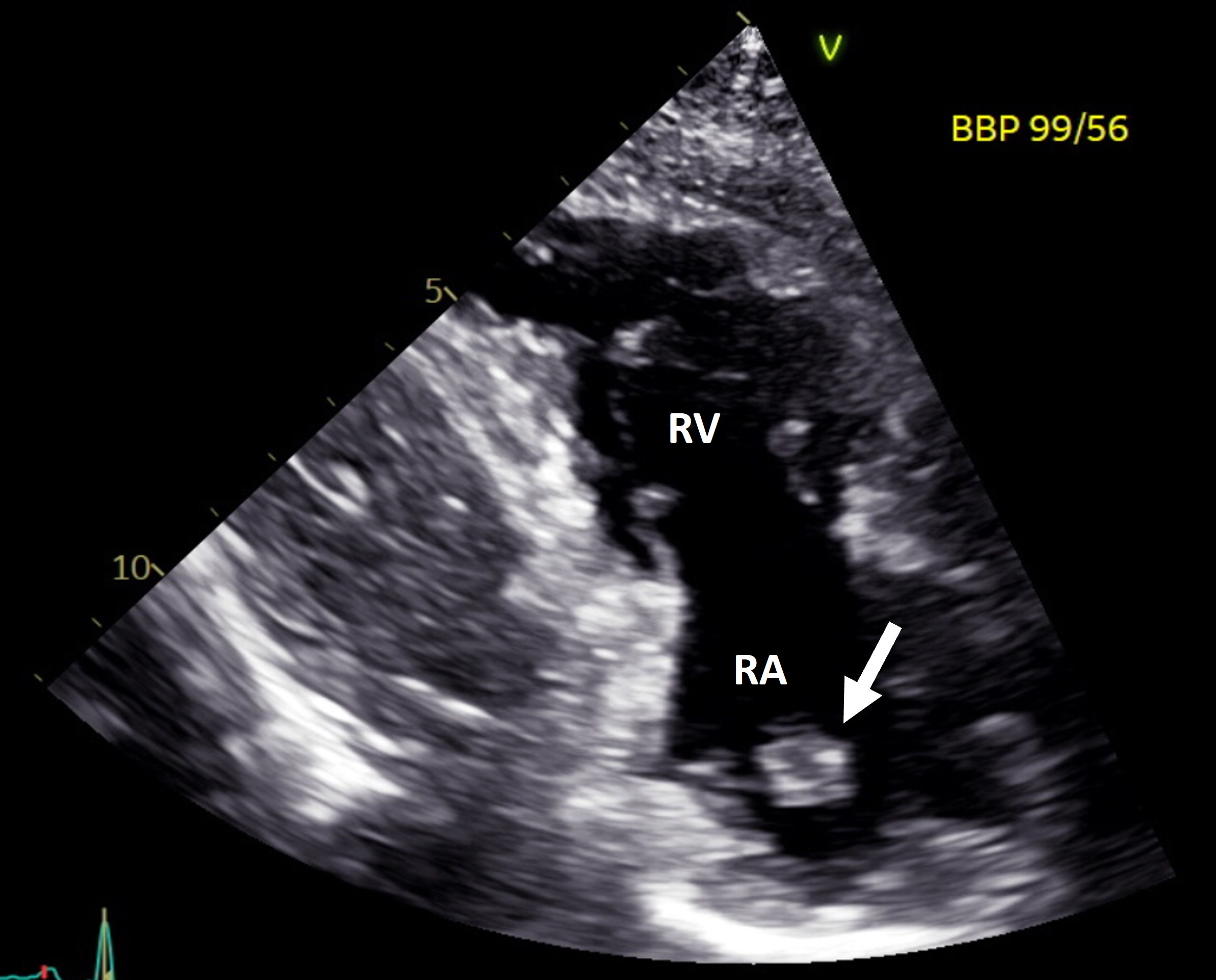
RA: right atrium, RV: right ventricle
Fig. 2. (A) TEE bi-caval view showing the ASA (white arrow); (B) Three-dimensional TEE view of the atrial septum from LA.
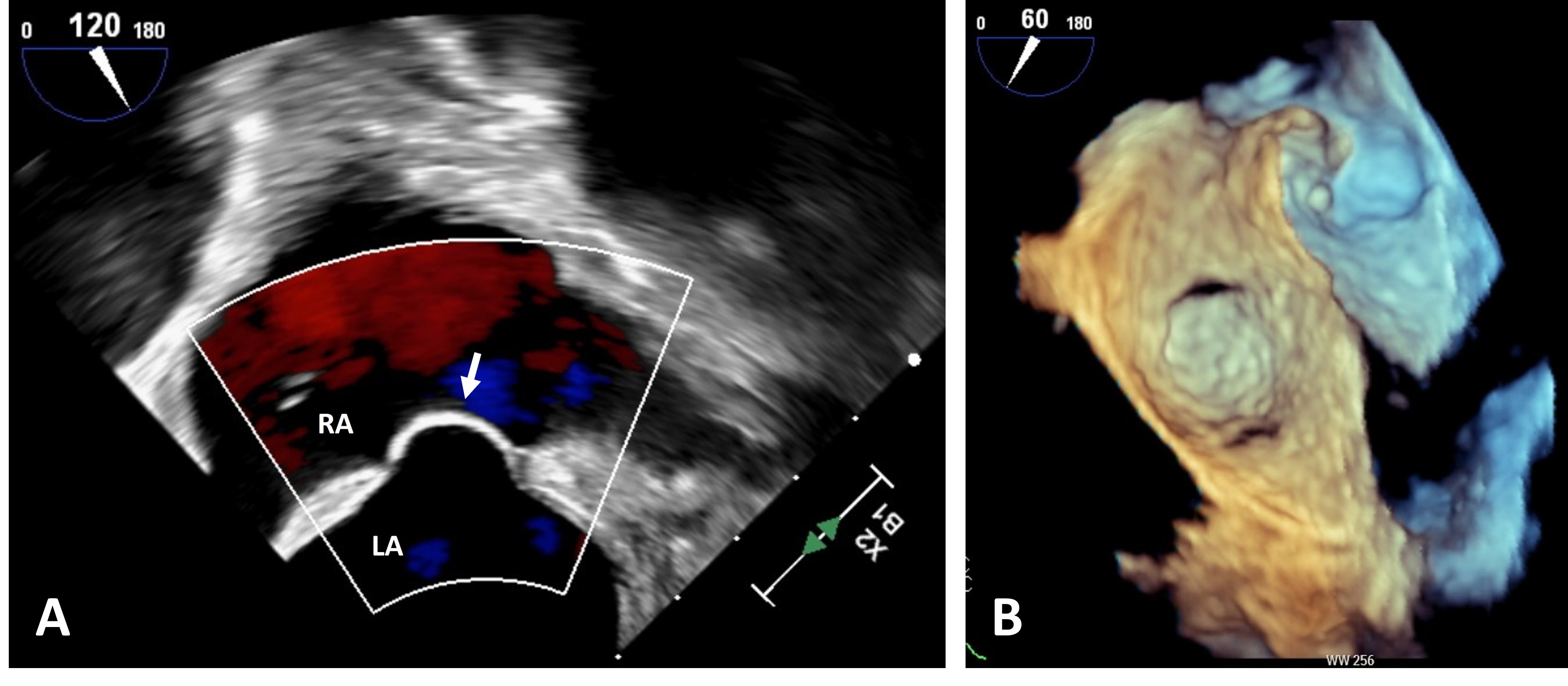
ASA: atrial septal aneurysm; LA: left atrium; RA: right atrium; TEE: transesophageal echocardiogram
Pseudomasses in the right atrium due to normal variants of benign structures such as the Eustachian/ Thebesian valve, Chiari network and crista terminalis are commonly seen in clinical practice. Due to the elevation plane and the mobility of ASA, ASA may masquerade as a pseudomass, not just in the right atrium, but possibly in the left atrium as well. Previous reports have described similar presentation of ASA as a mass in the right atrium.4,5,6
In addition to this atypical presentation, the authors have encountered 2 other atypical appearances of ASA—ASA may also present as a semi-circular structure in the right atrium in the right ventricle inflow view (Videos S5–6). The appearance of this structure resembles a winking eye. This is termed as an atypical appearance of the ASA as the winking sign. It can also manifest as a mobile linear echo in the LA suggestive of a “dissection flap” (Videos S7–8).
ASA has reported links with atrial arrhythmias, increased prevalence of cryptogenic stroke and other embolic events. Misdiagnosis of ASA can be minimised through a systematic echocardiographic approach. When a suspicious interatrial structure is visualised on TTE, acquiring multiple imaging planes—including subcostal, apical 4-chamber and right ventricular inflow views or orthogonal biplane imaging (X-plane)—can help clarify its attachment and motion pattern. The use of contrast echocardiography or agitated saline injection is particularly valuable in delineating the borders of the atrial septum and confirming the presence or absence of an interatrial shunt. In cases where TTE images are inconclusive, TEE provides superior spatial resolution and remains the gold standard for diagnosis, although it should be reserved as a last resort given its invasive nature.
Clinicians should be cognisant of these atypical appearances of ASA and employ an appropriate diagnostic strategy to avoid both misdiagnosis and unnecessary additional imaging.
Supplementary materials
Video S1. Transthoracic echocardiogram in the right ventricular inflow view showing a 0.9 cm x 1.4 cm well-circumscribed circular mass in the right atrium.
Video S2. Continuous acquisition of a number of cardiac cycles with the transducer rotating from basal parasternal short axis view to the right ventricular inflow view, showing that the “mass” is in fact an atrial septal aneurysm (ASA).
Video S3. Transesophageal echocardiogram bi-caval view of the atrial septum with the transducer at 120° showing the atrial septal aneurysm.
Video S4. Three-dimensional transesophageal echocardiogram view of the atrial septum from the left atrium.
Video S5. Transthoracic echocardiogram in the right ventricular inflow view showing a semi-circular structure in the right atrium (reproduced with permission from Yeo TC, Poh KK. Case-based comprehensive cardiology. Singapore: Springer Nature; to be published).
Video S6. Transthoracic echocardiogram in the apical 4-chamber view showing a large atrial septal aneurysm bulging into the right atrium (reproduced with permission from Yeo TC, Poh KK. Case-based comprehensive cardiology. Singapore: Springer Nature; to be published).
Video S7. Transthoracic echocardiogram in the parasternal long axis view showing a mobile linear echo suggestive of a “dissection flap” in the left atrium (reproduced with permission from Yeo TC, Poh KK. Case-based comprehensive cardiology. Singapore: Springer Nature; to be published).
Video S8. Transthoracic echocardiogram in the parasternal short axis view showing an atrial septal aneurysm bulging into the left atrium (Reproduced with permission from Yeo TC, Poh KK. Cas -based comprehensive cardiology. Singapore: Springer Nature; to be published).
REFERENCES
- Silvestry FE, Cohen MS, Armsby LB, et al. Guidelines for the Echocardiographic Assessment of Atrial Septal Defect and Patent Foramen Ovale: From the American Society of Echocardiography and Society for Cardiac Angiography and Interventions. J Am Soc Echocardiogr 2015;28:910-958.
- Gallet B, MC. Malergue, Adams C, et al. Atrial septal aneurysm–a potential cause of systemic embolism. An echocardiographic study. Br Heart J 1985;53:292-7.
- Olivares-Reyes A, Chan S, Lazar EJ, et al. Atrial septal aneurysm: A new classification in two hundred five adults. J Am Soc Echocardiogr 1997;10:644-56.
- Yeoh JK, Appelbe AF, Martin RP. Atrial septal aneurysm mimicking a right atrial mass on transesophageal echocardiography. Am J Cardiol 1991 68:827-8.
- Smith AJ, Panidis IP, Berger S, et al. Large atrial septal aneurysm mimicking a cystic right atrial mass. Am Heart J 1990;120:714-6.
- Ginon I. A closed interatrial septum aneurysm, filled with blood, mimicking a tumour in the right atrium. Eur J Echocardiogr 2000;1:289-90.
Not applicable as de-identified and anonymised data were used.
The author(s) declare there are no affiliations with or involvement in any organisation or entity with any financial interest in the subject matter or materials discussed in this manuscript.
Dr Ting Wei Teo, Department of Cardiology, National University Heart Centre Singapore, 5 Lower Kent Ridge Rd, Singapore 119074. Email: [email protected]
