

Dear Editor,
We are writing to present detailed findings of our recent study comparing digital polymerase chain reaction (dPCR) and quantitative polymerase chain reaction (qPCR) for the quantification of Epstein-Barr virus (EBV) deoxyribonucleic acid (DNA) in nasopharyngeal carcinoma (NPC). According to Global Cancer Observatory: Cancer Today 2020 statistics, NPC accounts for an estimated 129,000 new diagnoses and nearly 73,000 deaths annually,1 underscoring the need for accurate diagnostic and surveillance tools.
EBV is a major risk factor for NPC. While over 90% of the world’s population harbour latent EBV infections,2 the virus can persist in the nasopharyngeal epithelium and contribute to oncogenic mutations.3 EBV DNA and latent gene products are detectable in NPC and precancerous lesions, supporting its biological relevance.4 Persistently detectable plasma EBV DNA after treatment strongly predicts residual or recurrent disease, making serial EBV DNA monitoring an important component of patient follow-up.5
Surveillance after primary treatment remains challenging. Radiation-induced fibrosis, local oedema and post-treatment changes can obscure early recurrence on endoscopy or imaging, complicating decision-making.6 In this context, a sensitive biomarker like EBV DNA is essential for guiding critical treatment. Our study aimed to evaluate the diagnostic accuracy of dPCR for NPC detection and recurrence, and compare its performance with qPCR.
Although qPCR is the current standard for EBV DNA quantification, its reliance on calibration curves and reduced precision at low copy numbers may limit sensitivity for minimal residual disease (MRD) and early recurrence. dPCR allows absolute quantification and is expected to improve detection accuracy at low viral loads. We hypothesised that dPCR would outperform qPCR in plasma while recognising that any advantage might be reduced in serum. Accordingly, we compared the performance of dPCR versus qPCR across 2 EBV targets (BamHI-W and EBNA1) in both plasma and serum, and examined their potential clinical applications.
We conducted a case-control study at a single-centre tertiary university hospital, analysing 162 plasma and serum samples under institutional ethics approval. Two EBV gene targets were evaluated: the multi-copy BamHI-W sequence and the single-copy Epstein-Barr Nuclear Antigen 1 (EBNA1) gene.
The study comprised 2 cohorts. First, we prospectively collected 50 plasma samples from 38 NPC patients (stages I–IVA) and 12 healthy controls. Second, we analysed 112 archived serum samples from 28 patients with recurrent NPC and 28 matched healthy first-degree family members, with patient samples corresponding to pre-treatment, post-treatment, and recurrence. DNA was extracted using the Reliaprep Blood gDNA Miniprep System (Promega, Madison, WI, US). qPCR was performed on an Applied Biosystem StepOne Plus system (Applied Biosystems, Thermo Fisher Scientific, Waltham, MA, US). dPCR was performed on the Clarity dPCR system (JN Medsys, Singapore), following the manufacturer’s protocol. Diagnostic performance was assessed by receiver operating characteristic (ROC) analysis using the Youden J index (additional information for the clinical characteristics of the cohort and methods is in Supplementary Table S1).
The analysis of results from plasma samples showed superior performance of dPCR (Fig. 1). For BamHI-W, area under the curve (AUC) was 0.921 for dPCR compared to 0.865 for qPCR. For EBNA1, the difference was more marked (0.936 versus [vs] 0.792). Optimal cut-offs for each test were determined by maximising Youden J (Supplementary Table S2). At a cut-off of 31.5 copies/mL, plasma dPCR for BamHI-W achieved a sensitivity of 95.8% and specificity of 73.1%.
Fig. 1. Comparison of plasma EBV dPCR and qPCR in NPC: (A) BamHI-W levels across patient groups; (B) ROC curves for BamHI-W and EBNA1; and (C) Correlation of dPCR vs qPCR for both targets .
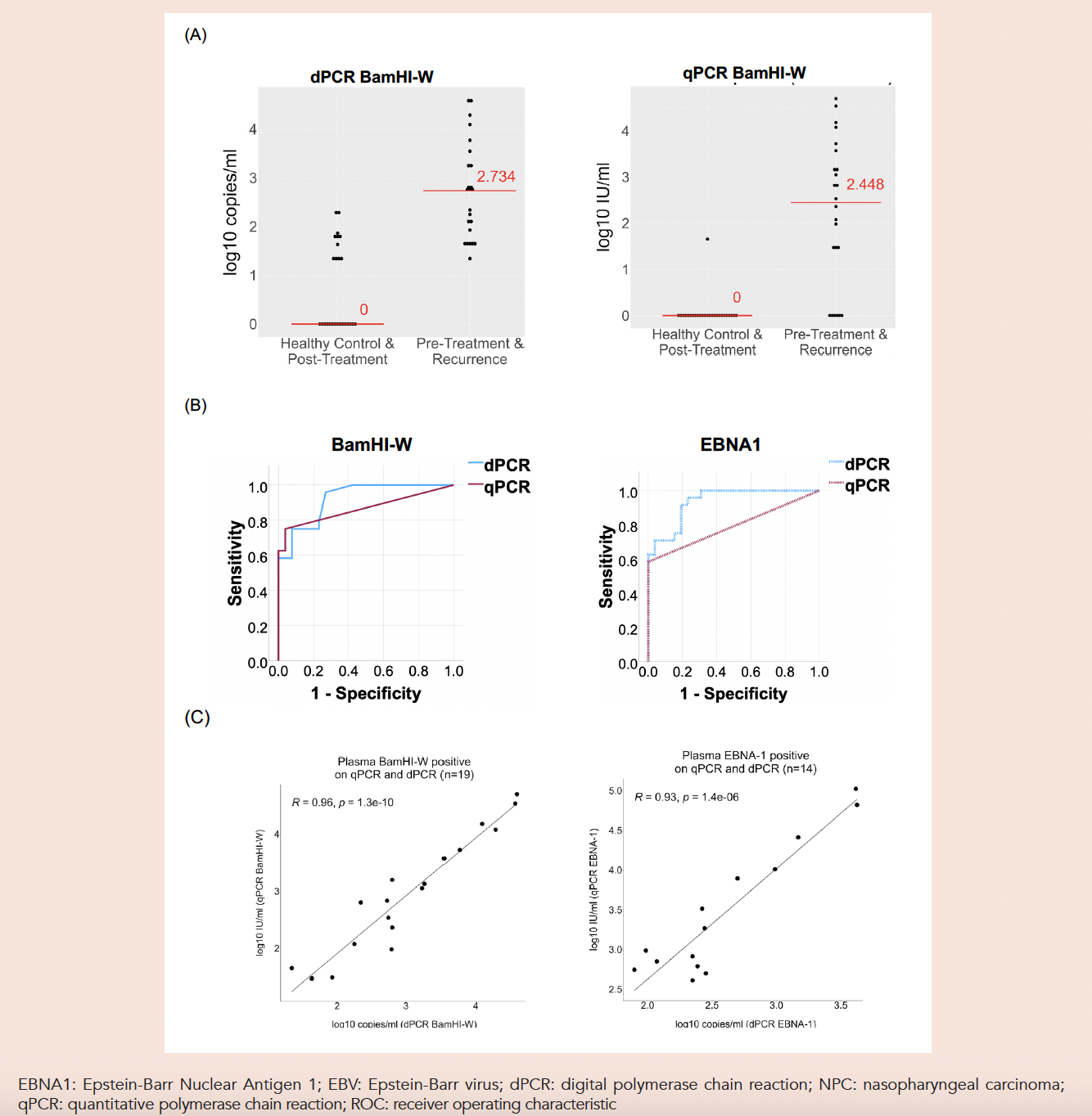
The EBNA1 findings were particularly notable. Despite being a single-copy target, EBNA1 showed strong discriminative performance on dPCR (cut-off 72 copies/mL: sensitivity 95.8% specificity 76.9%). This challenges the long-held belief that the multi-copy BamHI-W target is inherently superior for detection and suggests that absolute quantification may mitigate the analytical disadvantage of lower gene copy number.7
In contrast, serum analysis showed comparable performance between dPCR and qPCR (BamHI-W AUC 0.821 vs 0.832; EBNA1 AUC 0.683 vs 0.712; Supplementary Fig. S1). Patients with active disease had significantly higher median EBV DNA levels compared to healthy controls (Supplementary Figs. S3, S4). The reduced discriminability in serum is consistent with known differences in circulating DNA integrity and background genomic noise. This likely reflects a combination of biological and methodological factors: serum has lower tumour-derived DNA due to coagulation-associated degradation and greater release of leukocyte genomic DNA during clot formation, resulting in a lower proportion of tumour-specific DNA.8 Additional pre-analytical variables inherent to archived serum samples (including storage conditions and freeze–thaw cycles) may further reduce assay performance.8 Collectively, these factors may explain why dPCR displays a clear advantage in plasma but not in serum.
The superior performance of dPCR in plasma is attributable to its partitioning of samples into thousands of independent reactions, enabling absolute quantification without calibration curves, improving precision at low copy numbers. This facilitates detection of subtle changes in viral load that may be missed by qPCR.7
This work represents one of the first direct comparisons of dPCR and qPCR across 2 EBV targets in both plasma and serum. While plasma remains the preferred specimen for EBV DNA testing due to higher levels of tumour-derived DNA,9 serum findings retain clinical relevance in settings where it is routinely collected or when plasma samples are unavailable. Our longitudinal serum cohort also reaffirmed that patients with distant metastasis exhibit significantly higher EBV DNA loads than those with locoregional recurrence (P<0.01), highlighting EBV DNA’s value for characterising disease relapse.
Our findings have practical implications for clinical practice. Plasma should be the specimen of choice for EBV DNA quantification in NPC surveillance. dPCR may be particularly valuable in scenarios where sensitivity is critical: post-treatment monitoring, MRD assessment, and cases with equivocal radiological or endoscopic findings. Clinical trials evaluating treatment response with reproducibility and standardisation are crucial. EBNA1, despite being a single-copy gene, demonstrated strong performance on dPCR and warrants further evaluation as a routine clinical target.
In conclusion, our study demonstrates that plasma dPCR offers superior sensitivity and accuracy compared with qPCR for NPC detection and surveillance. Both BamHI-W and EBNA1 are useful targets, with EBNA1 showing unexpected potential in the dPCR setting. Earlier and more reliable identification of recurrence may allow more timely and potentially curative therapies. Further validation in larger prospective cohorts is needed to explore whether a combined BamHI-W/EBNA1 dPCR assay can provide even greater diagnostic power.
Supplementary materials
Supplementary Fig. S1. Comparison of serum EBV dPCR and qPCR in NPC.
Supplementary Fig. S2. Serum EBV DNA levels (EBNA1) in NPC patients (n=28) at 3 time points (pre-treatment, post-treatment and at recurrence) and healthy controls (n=28). Median values indicated by red lines.
Supplementary Fig. S3. Serum EBV DNA levels for NPC cases with recurrence, based on site of recurrent disease.
Supplementary Table S1. Clinical characteristics.
Supplementary Table S2. Performance of plasma EBV dPCR and qPCR.
Funding
This work was supported by the National Medical Research Council, Singapore, through the Clinician Scientist Award (CSAINV24jul-0011 to Joshua K. Tay) and the Large Collaborative Grant (OFLCG21jun-0013 to Kwok Seng Loh, Melvin Lee Kiang Chua and Jianjun Liu).
REFERENCES
- Bray F, Ferlay J, Soerjomataram I, et al. Global cancer statistics 2018: GLOBOCAN estimates of incidence and mortality worldwide for 36 cancers in 185 countries. CA Cancer J Clin 2018;68:394-424.
- Smatti MK, Al-Sadeq DW, Ali NH, et al. Epstein-barr virus epidemiology, serology, and genetic variability of LMP-1 oncogene among healthy population: An update. Front Oncol 2018;13;8:211.
- Farrell PJ. Epstein-Barr Virus and Cancer. Annu Rev Pathol Mech Dis 2019;14:29-53.
- Lee HM, Okuda KS, González FE, et al. Current Perspectives on Nasopharyngeal Carcinoma. In: Rhim, J., Dritschilo, A., Kremer, R. (eds) Human Cell Transformation. Advances in Experimental Medicine and Biology, vol 1164. Springer, Cham.
- Lin J-C, Wang W-Y, Chen KY, et al. Quantification of Plasma Epstein–Barr Virus DNA in Patients with Advanced Nasopharyngeal Carcinoma. N Engl J Med 2004;350:2461-70.
- Ak S, Kiliç C, Özlügedik S. Correlation of PET-CT, MRI and histopathology findings in the follow-up of patients with nasopharyngeal cancer. Braz J Otorhinolaryngol 2020;87:643-8.
- Bio-Rad.com. Digital PCR and Real-Time PCR (qPCR) Choices for Different Applications. https://www.bio-rad.com/en-sg/applications-technologies/digital-pcr-real-time-pcr-qpcr-choices-for-different-applications?ID=OENHBB15. Accessed 17 April 2021.
- Stevens SJC, Pronk I, Middeldorp JM. Toward standardization of Epstein-Barr virus DNA load monitoring: Unfractionated whole blood as preferred clinical specimen. J Clin Microbiol 2001;39:1211-6.
- Liu Y, Fang Z, Liu L, et al. Detection of Epstein-Barr Virus DNA in Serum or Plasma for Nasopharyngeal Cancer: A Meta-Analysis. Genet Test Mol Biomarkers 2011;15:495-502.
This study was conducted in accordance with the Declaration of Helsinki and received formal approval from the National Healthcare Group Domain Specific Review Board (2006/00149). Written informed consent was obtained from all participants prior to their inclusion in the study. Use of plasma sample was approved by National Health Group Domain Specific Review Board (2016/00045).
Johnson Ng is founder of JN Medsys, the manufacturer of the dPCR platform used in this study. All data interpretation was independent of Johnson Ng. All other authors declare they have no affiliations or financial involvement with any commercial organisation with a direct financial interest in the subject or materials discussed in the manuscript.
Dr Joshua K Tay, Department of Otolaryngology–Head & Neck Surgery, National University of Singapore, 1E Kent Ridge Road, Level 7, Singapore 119228. Email: [email protected]; Dr Benedict Yan, Department of Laboratory Medicine, National University Hospital, Singapore, 5 Lower Kent Ridge Road, Level 7, Singapore 1190074. Email: [email protected]
