

ABSTRACT
Introduction: This study aimed to compare the clinical outcomes of endoscopic combined intrarenal surgery (ECIRS) in the prone hip-flexed split-leg position with conventional percutaneous nephrolithotomy (PCNL) for the treatment of complex renal calculi.
Methods: We retrospectively analysed 168 patients with complex renal calculi, including 82 who underwent ECIRS and 86 who underwent conventional PCNL. Baseline demographic and stone characteristics, perioperative outcomes, initial stone-free rate (SFR) within 1–3 days after surgery, final SFR after completion of all planned treatments, auxiliary procedures, double-J stent indwelling duration, length of hospital stay, and postoperative complications were compared between groups. Logistic regression analyses were performed to evaluate factors associated with residual stones after the initial procedure, with additional clinically informed sensitivity analysis using prespecified covariates.
Results: Baseline demographic characteristics, stone features, and postoperative outcomes, including intraoperative ureteral catheterization time, complication rates, Clavien–Dindo classification, and final SFR after completion of all planned treatments, were comparable between the two groups (all P>0.05). In this retrospective cohort, ECIRS in the prone hip-flexed split-leg position was associated with earlier stone clearance after the index procedure, reflected by a higher initial SFR (74.39% versus [vs] 50.00%, P=0.001) and a lower requirement for auxiliary procedures (29.27% vs 46.51%, P=0.021). These findings were accompanied by a shorter hospital stay (6.44 vs 8.61 days, P<0.001), shorter double-J stent indwelling duration (2.81 vs 4.79 weeks, P=0.002), and shorter operative time (P=0.012). Multivariable and sensitivity analyses further demonstrated that ECIRS was independently associated with lower odds of residual stones after the initial procedure (odds ratio 0.40, 95% confidence interval 0.20–0.80, P=0.009).
Conclusion: Single-position ECIRS in the prone hip-flexed split-leg configuration was feasible for selected patients with complex renal calculi and was associated with earlier stone clearance and shorter perioperative recovery metrics. However, the final SFR after completion of all planned treatments was comparable between groups, and whether this positional configuration confers benefit beyond ECIRS in alternative positions could not be determined from this comparison. Further prospective studies are required to validate these findings.
CLINICAL IMPACT
What is New
- This study showed that ECIRS in the modified prone hip-flexed split-leg position was associated with a significantly higher initial stone-free rate (74.39% versus [vs] 50.00%), shorter hospital stay (6.44 vs 8.61 days), and shorter double-J stent indwelling time (2.81 vs 4.79 weeks) compared with conventional PCNL for complex renal calculi, without an associated increase in complication rates.
- The modified position eliminates the need for intraoperative repositioning, optimising surgical access and efficiency.
Clinical Implications
- Our findings suggest that ECIRS in the prone hip-flexed split-leg position may represent a feasible alternative for selected patients with complex renal calculi in centres with established endourological infrastructure.
- This approach was associated with earlier stone clearance and shorter perioperative recovery metrics; however, residual confounding inherent to the retrospective study design precludes causal interpretation of these associations.
Renal calculi are among the most prevalent urological diseases worldwide, severely impairing patients’ quality of life and increasing the healthcare burden. Epidemiological data from China showed that the national prevalence of renal calculi was 6.4% in 2013–2014,1 and the hospitalisation-related healthcare burden has continued to increase in recent years.2 Among these, complex renal stones—such as staghorn calculi, multiple stones, or stones associated with anatomical abnormalities—pose substantial clinical challenges due to their high recurrence rate, treatment difficulty, and increased risk of complications. These cases require efficient and safe treatment strategies.
Percutaneous nephrolithotomy (PCNL) is regarded as the gold standard for treating complex renal stones, enabling stone fragmentation and removal through a percutaneous tract.3 However, in the presence of multiple complex calculi, PCNL may require multiple tracts or repeated procedures, which can be associated with greater trauma, delayed recovery, and higher complication risks. In addition, the conventional prone position has inherent limitations, including the need for patient repositioning and technical constraints when simultaneous retrograde ureteroscopic access is required.4
Endoscopic combined intrarenal surgery (ECIRS) integrates the advantages of percutaneous nephroscopy and ureteroscopy, facilitating stone fragmentation and fragment retrieval through dual access. Since its introduction with the Galdakao-modified supine Valdivia position in 2008,5 ECIRS has undergone multiple modifications in positioning techniques, including the reverse lithotomy position, prone hip-flexed split-leg position, and knee-chest prone position. These innovations have expanded positioning strategies for ECIRS; however, adoption may still be influenced by surgeon familiarity, equipment availability, and institutional workflow.
To facilitate simultaneous antegrade and retrograde access during ECIRS, we adopted a prone hip-flexed split-leg position that allows the procedure to be completed without intraoperative repositioning. The present study aimed to evaluate the feasibility and perioperative outcomes of ECIRS performed in this position compared with conventional PCNL for the treatment of complex renal calculi.
METHODS
Study design
A retrospective cohort analysis was conducted on the clinical data of patients diagnosed with complex renal stones at Jinhua Hospital, Zhejiang University School of Medicine, between May 2022 and July 2024. Patients who underwent either PCNL or ECIRS for stone removal were included. The inclusion criteria were as follows: (1) age ≥18 years; (2) preoperative confirmation of complex renal stones by ultrasonography and computed tomography (CT), defined as stones meeting any of the following criteria: (a) a maximum diameter >2 cm; (b) staghorn calculi; or (c) multiple renal stones (≥2 stones) with a S.T.O.N.E. score ≥7 points6; (3) patients treated with ECIRS in the prone hip-flexed split-leg position or with traditional PCNL; and (4) complete clinical data available. The exclusion criteria were as follows: (1) anatomical abnormalities of the kidney or functional/anatomical absence of a unilateral kidney; (2) coagulation disorders or a history of prior PCNL; (3) concomitant active urinary tract infection uncontrolled after standard preoperative treatment, tuberculosis, or severe cardiopulmonary dysfunction; and (4) incomplete clinical data compromising data integrity assessment. The distribution of patients meeting each criterion for complex renal stones is provided in Supplementary Table S1. Cases with missing key variables were excluded from the final analysis, and no missing data imputation was performed.
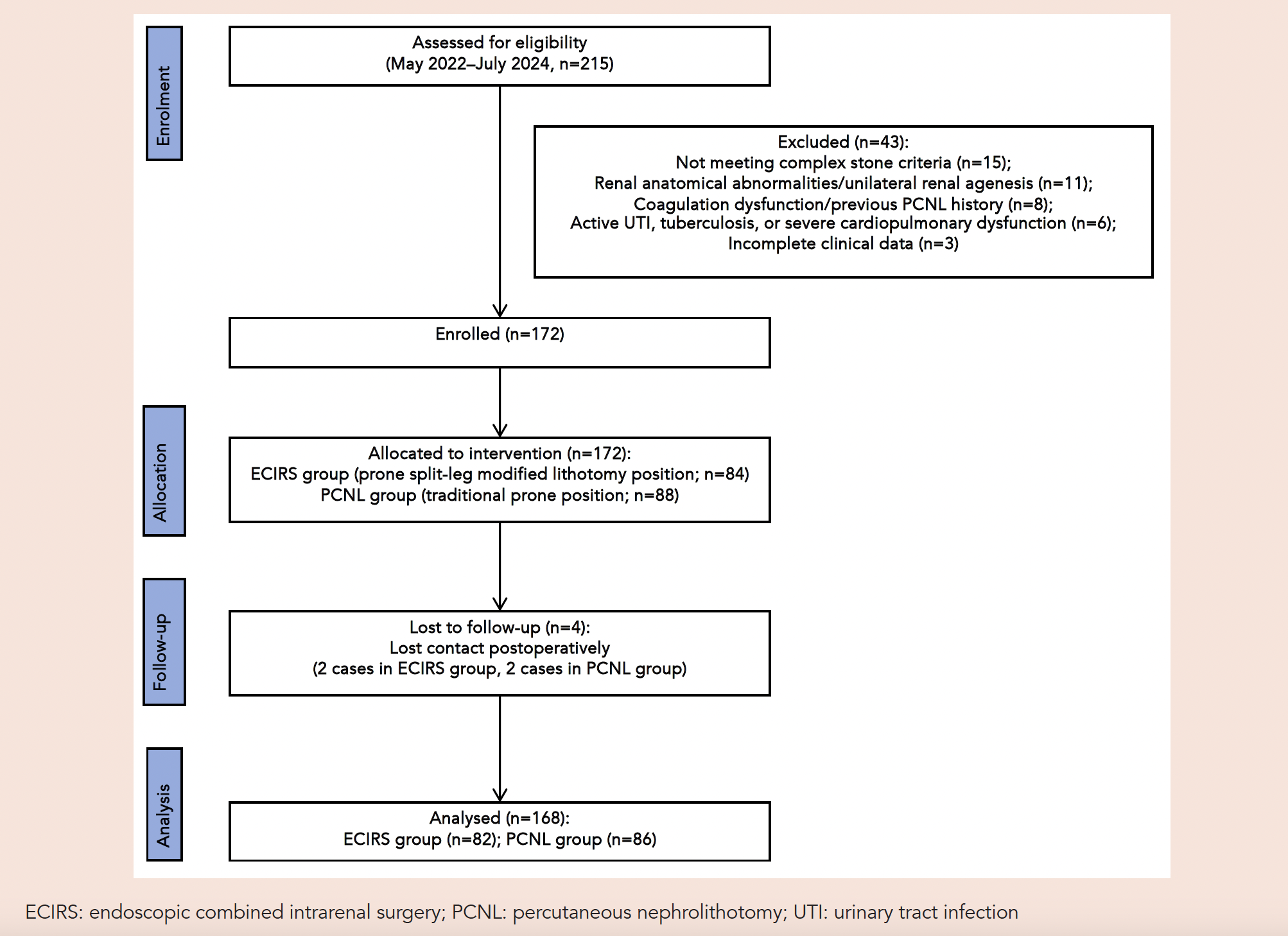
This study was approved by the respective ethics committee. Written informed consent was obtained from all enrolled patients. The patient grouping was determined through comprehensive evaluation by 3 experienced urologists, based on the S.T.O.N.E. score quantifying stone complexity, renal anatomical characteristics, and the patient’s informed consent. The detailed process of patient screening, inclusion, exclusion, and final grouping is illustrated in Fig. 1.
Fig. 1. Flow diagram of patient screening.
Preoperative evaluation
All patients underwent standardised preoperative preparation, including detailed medical history collection, physical examination, routine laboratory tests, and imaging evaluation. Preoperative workup included urinalysis, urine culture, and antibiotic susceptibility testing. For patients with urinary tract infection, targeted antibiotic therapy was administered preferentially according to urine culture and susceptibility test results. In the absence of definitive susceptibility test results, empirical antibiotics, primarily cephalosporins, and selected quinolones based on local antimicrobial susceptibility patterns were administered under antimicrobial stewardship principles; and the use of broad-spectrum antibiotics was strictly controlled to avoid the development of drug resistance. For patients with confirmed preoperative urinary tract infection, targeted anti-infective therapy was continued for 7 days, followed by repeat urine culture and urinalysis before surgery. Patients were included in the surgical protocol only when the urine culture result turned negative, the white blood cell count in urinalysis returned to normal, and the infection was confirmed to be effectively controlled. Postoperative antibiotic therapy was administered according to perioperative infection status, urine culture results, and institutional antimicrobial stewardship protocols. The complexity of renal stones was objectively assessed using the S.T.O.N.E. scoring system,7 based on non-contrast CT images. This scoring system comprises 5 parameters: S (stone size), T (tract length), O (obstruction), N (number of involved calyces), and E (essence/Hounsfield units). The sum of these 5 items yielded a total score ranging from 5 to 13.
Surgical technique
Percutaneous nephrolithotomy
Patients in the PCNL group underwent the conventional 2-step puncture method. Under general anaesthesia in the lithotomy position, a 5 Fr open-ended ureteral catheter was inserted into the upper urinary tract and an 18 Fr Foley catheter into the bladder. The patient was then repositioned to the traditional prone position, with pressure points (chest, iliac crest, and knees) adequately protected. Retrograde pyelography was subsequently performed, and renal calyx puncture was achieved under fluoroscopic and ultrasonographic guidance. A 0.035-inch stiff guidewire was advanced and coiled within the renal pelvis or ureter to ensure stability. Serial fascial dilators (16 Fr, 18 Fr, or 24 Fr) were used to sequentially establish a 16 Fr, 18 Fr, or 24 Fr percutaneous tract, followed by placement of a corresponding working sheath. A 20 Fr nephroscope was typically introduced through the standard 24 Fr tract, whereas an appropriately sized endoscope was selected for smaller-calibre tracts. Lithotripsy and stone extraction were then performed under direct endoscopic vision.
Endoscopic combined intrarenal surgery
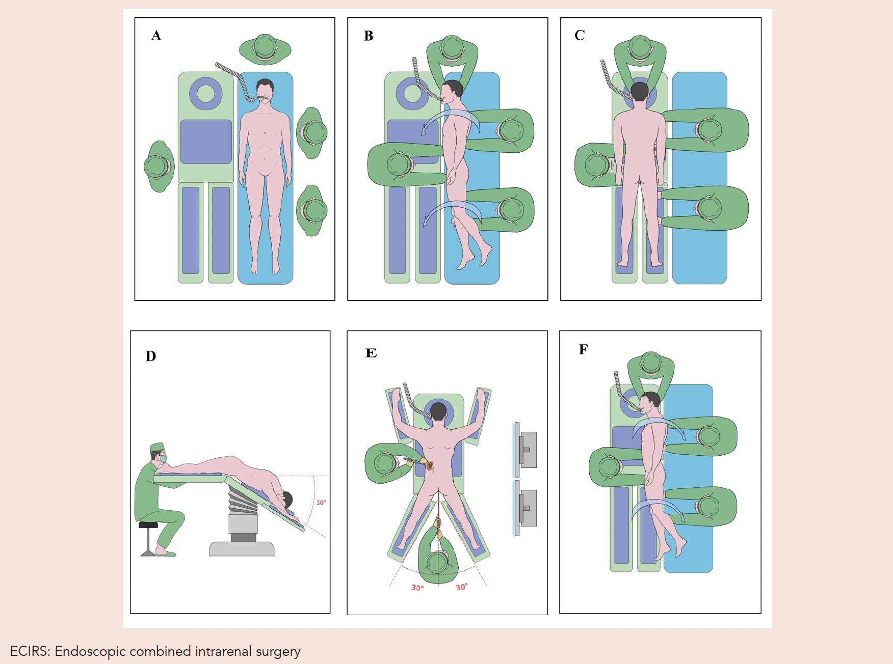
After induction of general anaesthesia, patients were transferred from the transport bed to the operating table in a coordinated team effort and placed in the prone hip-flexed split-leg position. Strategic padding was applied at pressure points (chest, iliac crest, and knees) to prevent neurovascular injury. Both lower limbs were positioned on padded supports aligned with the operating table, with approximately 20-degree hip flexion and an inter-leg distance of at least 30 cm. The operating table was raised by ~30 cm, the head was tilted downwards, and the foot end was elevated by 30 cm, resulting in a perineal elevation of 60 degrees. The procedure began with cystoscopy, followed by retrograde insertion of a 5–7 Fr open-ended ureteral catheter into the renal pelvis to create artificial hydronephrosis, after which a 12–14 Fr ureteral access sheath was advanced over a 0.035-inch guidewire. The operating table was then levelled, and a 7.5 Fr flexible ureteroscope was inserted. Under combined ultrasound and flexible ureteroscopic guidance, a percutaneous renal tract was established using serial fascial dilators and corresponding Amplatz sheaths (16 Fr, 18 Fr, or 24 Fr), selected according to stone burden, calyceal anatomy, and intraoperative surgical requirements. Large stones were fragmented with an ultrasonic/pneumatic lithotripter via nephroscopy, while small stones in difficult-to-access locations were treated with holmium laser through the flexible ureteroscope. When patients were transferred to the recovery bed, the same position was maintained (20-degree hip flexion and ≥30 cm inter-leg distance), thus avoiding intraoperative repositioning (Fig. 2).
Fig. 2. Steps of establishing the prone hip-flexed split-leg position for ECIRS. (A) The patient receives anaesthesia in the supine position on the transport bed prior to transfer. (B) The patient is turned to the prone position on the adjacent operating table with coordinated team assistance. (C) Strategic padding is applied at pressure points (chest, iliac crest, and knees) to prevent neurovascular injury. (D) Retrograde ureteral catheter insertion to establish artificial hydronephrosis prior to percutaneous puncture. (E) Simultaneous manipulation of percutaneous nephroscope (upper image) and transurethral ureteroscope (lower image). (F) Patient transferred to the recovery bed while maintaining the prone hip-flexed split-leg alignment.
All procedures were performed by three experienced urologists. A ureteral catheter was routinely left indwelling in all patients, and a 6 Fr double-J ureteral stent was placed strictly according to unified objective criteria in patients with intraoperatively confirmed significant ureteral oedema, residual stone fragments ≥2 mm with potential risk of postoperative obstruction or requiring subsequent auxiliary treatment, or anticipated need for subsequent intervention. Stent removal was performed by a dedicated urological team after resolution of gross haematuria and flank pain, adequate control of urinary tract infection confirmed by urinalysis, and imaging confirmation of no significant residual calculi or urinary tract obstruction. The ureteral catheter and Foley catheter were removed after confirmation of no urinary extravasation, haematuria, or other abnormalities following evaluation at 24–48 hours postoperatively. Operative time was defined as the interval from the initiation of cystoscopy to the completion of Foley catheter and double-J ureteral stent placement. Length of hospital stay was defined as the duration from patient admission to discharge.
Postoperative assessment
Serum haemoglobin and creatinine levels were measured within 24 hours after surgery and compared with preoperative values. Abdominal CT was performed 1–3 days postoperatively to assess residual stones and determine the initial SFR. CT urography was reserved for patients with severe haemorrhagic complications or when residual stones could not be accurately evaluated on standard postoperative CT. Stone-free status was defined as the absence of residual intrarenal stone fragments ≥4 mm on postoperative imaging.8 For patients with intrarenal residual stones ≥4 mm detected on postoperative imaging, the need for adjuvant intervention was determined comprehensively based on the patients’ clinical symptoms and the size and location of the residual stones. Adjuvant interventions included extracorporeal shock wave lithotripsy, secondary percutaneous nephrolithotomy, and flexible ureteroscopic lithotripsy. Postoperative complications were graded according to the Clavien–Dindo classification system9: Grade I complications required no specific intervention or symptomatic management only, and included low-grade postoperative fever and mild haematuria, which were managed with fluid support, close vital sign monitoring, and symptomatic treatment. Grade II complications required pharmacological intervention and included febrile infection and minor leakage around the nephrostomy tube. Targeted antimicrobial therapy was administered according to urine culture and antimicrobial susceptibility results, and nephrostomy tube leakage was managed with local disinfection and sterile dressing changes. Grade III complications required invasive intervention and included severe haemorrhage, perirenal haematoma, ureteral mucosal injury, and severe infection. Corresponding treatments included endovascular embolisation for haemostasis, ultrasound-guided percutaneous drainage of hematoma, placement of a 6 Fr double-J ureteral stent for ureteral support and healing, and intensive anti-infective therapy with broad-spectrum antibiotics, with additional anaerobic coverage when necessary. Follow-up imaging was performed 3 months after completion of all planned treatments, using kidney-ureter-bladder (KUB) radiography and/or CT, to determine the final SFR. Final stone-free status was defined as the absence of residual fragments ≥4 mm or the presence of clinically insignificant residual fragments not requiring further intervention.
Statistical analysis
Continuous variables were expressed as mean ± standard deviation or median (interquartile range). Group comparisons were performed using Student’s t-test or the Mann-Whitney U test. Categorical variables were expressed as frequencies (percentages) and compared using the chi-square test or Fisher’s exact test. Univariate and multivariable logistic regression analyses were performed to identify factors associated with residual stones after the initial procedure. To reduce potential confounding bias inherent to retrospective analyses, additional clinically informed sensitivity analyses were performed using prespecified clinically relevant covariates. All statistical analyses were conducted using R software version 4.3.1 (R Core Team, Vienna, Austria), and a two-tailed P<0.05 was considered statistically significant.
RESULTS
Baseline characteristics of patients
A total of 168 patients with complex renal stones were included, of whom 86 underwent PCNL and 82 underwent ECIRS. No significant differences were observed in baseline demographic or stone characteristics between the 2 groups (all P>0.05, Table 1). The mean S.T.O.N.E. score was 8.1 ± 1.1 in the ECIRS group and 8.3 ± 0.9 in the PCNL group, indicating comparable stone complexity between groups.
Table 1. Baseline demographic and stone characteristics of patients.
|
Variables |
Total (n=168) |
PCNL group (n=86) |
ECIRS group (n=82) |
P |
|
Age (years) |
60.17 ± 11.41 |
59.59 ± 12.39 |
60.79 ± 10.32 |
0.495 |
|
BMI (kg/m²) |
24.50 ± 3.99 |
25.02 ± 4.10 |
23.93 ± 3.79 |
0.075 |
|
Sex, n (%) |
|
|
|
0.388 |
|
Female |
65 (38.69) |
36 (41.86) |
29 (35.37) |
|
|
Male |
103 (61.31) |
50 (58.14) |
53 (64.63) |
|
|
Stone side, n (%) |
|
|
|
0.769 |
|
Right |
80 (47.62) |
40 (46.51) |
40 (48.78) |
|
|
Left |
88 (52.38) |
46 (53.49) |
42 (51.22) |
|
|
S.T.O.N.E. score |
8.2 ± 1.0 |
8.3 ± 0.9 |
8.1 ± 1.1 |
0.159 |
|
S score |
1.4 ± 0.5 |
1.4 ± 0.5 |
1.4 ± 0.6 |
0.979 |
|
T score |
1.3 ± 0.5 |
1.3 ± 0.5 |
1.3 ± 0.5 |
0.659 |
|
O score |
1.5 ± 0.5 |
1.5 ± 0.5 |
1.5 ± 0.5 |
0.709 |
|
N score |
2.2 ± 0.6 |
2.3 ± 0.6 |
2.2 ± 0.7 |
0.276 |
|
E score |
1.8 ± 0.4 |
1.8 ± 0.3 |
1.7 ± 0.4 |
0.407 |
|
Preoperative haemoglobin (g/L) |
135.49 ± 17.52 |
136.72 ± 18.30 |
134.20 ± 16.67 |
0.351 |
|
Preoperative creatinine (μmol/L) |
103.63 ± 33.18 |
101.40 ± 33.92 |
105.98 ± 32.42 |
0.372 |
BMI: body mass index; ECIRS: endoscopic combined intrarenal surgery; PCNL: percutaneous nephrolithotomy; S.T.O.N.E.: stone size, tract length, obstruction, number of involved calyces, and essence
In the S.T.O.N.E. nephrolithometry score, S represents stone size; T, tract length; O, degree of obstruction; N, number of involved calyces; and E, stone essence or stone density. Values for the total S.T.O.N.E. score and its components are presented as means of integer-based scores.
Surgical and postoperative outcomes
According to the surgical data presented in Table 2, no significant differences were observed between the two groups in intraoperative ureteral catheterisation time, haemoglobin change, complication rate, or Clavien–Dindo classification. The operative time was shorter in the ECIRS group than in the PCNL group (102.38 ± 27.64 min vs 114.31 ± 33.18 min, P=0.012). Postoperative serum creatinine was lower in the ECIRS group (89.96 ± 21.68 μmol/L vs 96.78 ± 18.27 μmol/L, P=0.028), and creatinine change also differed between groups (median interquartile range [IQR]: ECIRS, −8.92 [−37.34 to 9.68] vs PCNL, 2.22 [−28.05 to 17.79], P=0.025). The double-J stent indwelling duration was likewise shorter in the ECIRS group than in the PCNL group (median IQR: 2.81 [2.04 to 3.88] weeks vs 4.79 [1.64 to 10.73] weeks, P=0.002).
The ECIRS group had a shorter hospital stay than the PCNL group (6.44 ± 1.98 days vs 8.61 ± 3.37 days, P<0.001). Regarding stone clearance, ECIRS was associated with a higher initial SFR within 1–3 days after the index procedure (74.39% [61/82] vs 50.00% [43/86], P=0.001) and a lower rate of auxiliary procedures (29.27% vs 46.51%, P=0.021). Specifically, 24 patients in the ECIRS group required additional procedures after the initial treatment, compared with 40 patients in the PCNL group. However, the final SFR at 3 months after completion of all planned treatments did not differ significantly between the 2 groups (95.12% [78/82] vs 93.02% [80/86], P=0.804).
Table 2. Surgical and postoperative characteristics.
|
Variables |
Total (n=168) |
PCNL group (n=86) |
ECIRS group (n=82) |
P |
|
Postoperative haemoglobin (g/L) |
129.42 ± 21.05 |
130.18 ± 19.76 |
128.62 ± 22.41 |
0.631 |
|
Postoperative creatinine (μmol/L) |
93.45 ± 20.24 |
96.78 ± 18.27 |
89.96 ± 21.68 |
0.028 |
|
Operative time (min) |
108.48 ± 31.09 |
114.31 ± 33.18 |
102.38 ± 27.64 |
0.012 |
|
Intraoperative ureteral catheterisation time (min) |
7.05 ± 2.53 |
6.95 ± 2.59 |
7.16 ± 2.49 |
0.601 |
|
Hospital stay (days) |
7.55 ± 2.98 |
8.61 ± 3.37 |
6.44 ± 1.98 |
<0.001 |
|
Haemoglobin change (postoperative − preoperative, g/L) |
−4.48 (−13.68 to 2.38) |
−7.92 (−14.10 to −0.49) |
−1.95 (−12.54 to 3.29) |
0.071 |
|
Creatinine change (postoperative − preoperative, μmol/L) |
−3.35 (−36.65 to 14.74) |
2.22 (−28.05 to 17.79) |
−8.92 (−37.34 to 9.68) |
0.025 |
|
Double-J stent placement, n (%) |
|
|
|
0.900 |
|
No |
24 (14.29) |
12 (13.95) |
12 (14.63) |
|
|
Yes |
144 (85.71) |
74 (86.05) |
70 (85.37) |
|
|
Double-J stent indwelling duration (weeks) |
3.40 (2.00, 6.19) |
4.79 (1.64, 10.73) |
2.81 (2.04, 3.88) |
0.002 |
|
Complications, n (%) |
|
|
|
0.952 |
|
No |
100 (59.52) |
51 (59.30) |
49 (59.76) |
|
|
Yes |
68 (40.48) |
35 (40.70) |
33 (40.24) |
|
|
Clavien-Dindo grade, n (%) |
|
|
|
0.621 |
|
No |
100 (59.52) |
51 (59.30) |
49 (59.76) |
|
|
I |
40 (23.81) |
18 (20.93) |
22 (26.83) |
|
|
II |
14 (8.33) |
8 (9.30) |
6 (7.32) |
|
|
III |
14 (8.33) |
9 (10.47) |
5 (6.10) |
|
|
Initial SFR, n (%) |
|
|
|
0.001 |
|
Stone-free |
104 (61.90) |
43 (50.00) |
61 (74.39) |
|
|
Residual stone |
64 (38.10) |
43 (50.00) |
21 (25.61) |
|
|
Final SFR, n (%) |
|
|
|
0.804 |
|
Stone-free |
158 (94.05) |
80 (93.02) |
78 (95.12) |
|
|
Residual stone |
10 (5.95) |
6 (6.98) |
4 (4.88) |
|
|
Auxiliary treatment, n (%) |
|
|
|
0.021 |
|
No |
104 (61.90) |
46 (53.49) |
58 (70.73) |
|
|
Yes |
64 (38.10) |
40 (46.51) |
24 (29.27) |
|
ECIRS: endoscopic combined intrarenal surgery; PCNL: percutaneous nephrolithotomy; SFR: stone-free rate
Continuous variables are presented as mean ± standard deviation (SD) or median (interquartile range [IQR]), as appropriate. For variables presented as median (IQR), the values in parentheses represent the 25th and 75th percentiles. Haemoglobin and creatinine changes were calculated as postoperative values minus preoperative values. Intraoperative ureteral catheterisation time refers to the time required for retrograde ureteral catheter placement during the procedure. Double-J stent indwelling duration was calculated among patients who received double-J stent placement.
Factors associated with initial SFR
Following the first treatment, residual stones were observed in 43 patients (50.00%) in the PCNL group and in 21 patients (25.61%) in the ECIRS group. To further explore factors associated with initial stone-free status, patients were stratified according to whether they achieved stone-free status after the initial procedure. As shown in Table 3, significant differences were observed between the stone-free and residual-stone groups in age, body mass index (BMI), S.T.O.N.E. score, operative time, and operative approach (all P<0.05).
Table 3. Clinical and perioperative characteristics stratified by initial stone-free status.
|
Variables |
Stone-free (n=104) |
Residual stone (n=64) |
P |
|
Age (years) |
58.77 ± 10.82 |
62.45 ± 12.04 |
0.042 |
|
BMI (kg/m²) |
23.97 ± 3.36 |
25.32 ± 4.72 |
0.049 |
|
S.T.O.N.E. score |
8.0 ± 1.0 |
8.4 ± 1.1 |
0.019 |
|
Preoperative haemoglobin (g/L) |
134.64 ± 16.77 |
136.87 ± 18.73 |
0.425 |
|
Postoperative haemoglobin (g/L) |
129.19 ± 18.87 |
129.79 ± 24.32 |
0.868 |
|
Preoperative creatinine (μmol/L) |
101.84 ± 32.35 |
106.55 ± 34.53 |
0.373 |
|
Postoperative creatinine (μmol/L) |
92.68 ± 21.31 |
94.71 ± 18.45 |
0.529 |
|
Operative time (min) |
104.72 ± 29.98 |
114.60 ± 32.13 |
0.045 |
|
Haemoglobin change (postoperative − preoperative, g/L) |
−4.48 (−12.64 to 1.84) |
−4.44 (−14.31 to 3.31) |
0.660 |
|
Creatinine change (postoperative − preoperative, μmol/L) |
−1.21 (−36.65 to 15.95) |
−5.16 (−34.55 to 11.33) |
0.564 |
|
Sex, n (%) |
|
|
0.088 |
|
Female |
35 (33.65) |
30 (46.88) |
|
|
Male |
69 (66.35) |
34 (53.12) |
|
|
Stone side, n (%) |
|
|
0.880 |
|
Right |
50 (48.08) |
30 (46.88) |
|
|
Left |
54 (51.92) |
34 (53.12) |
|
|
Complications, n (%) |
|
|
0.057 |
|
No |
69 (66.35) |
33 (51.56) |
|
|
Yes |
35 (33.65) |
31 (48.44) |
|
|
Operative approach, n (%) |
|
|
0.001 |
|
PCNL |
43 (41.35) |
43 (67.19) |
|
|
ECIRS |
61 (58.65) |
21 (32.81) |
|
BMI: body mass index; ECIRS: endoscopic combined intrarenal surgery; PCNL: percutaneous nephrolithotomy; S.T.O.N.E.: stone size, tract length, obstruction, number of involved calyces, and essence
Continuous variables are presented as mean ± standard deviation (SD) or median (interquartile range [IQR]), as appropriate. For variables presented as median (IQR), the values in parentheses represent the 25th and 75th percentiles.
Subsequent univariate logistic regression analysis (Table 4) showed that surgical modality, age, BMI, S.T.O.N.E. score, and operative time were associated with the risk of residual stones after the initial procedure (all P<0.05). Variables with statistical significance in the univariate analysis were subsequently entered into the primary multivariable logistic regression model. Surgical modality, S.T.O.N.E. score, and age remained independently associated with residual stones in the primary multivariable analysis (all P<0.05). To further evaluate the robustness of the findings, an additional clinically informed multivariable model—incorporating prespecified clinically relevant covariates, including age, sex, BMI, S.T.O.N.E. score, stone side, and complication occurrence—was subsequently constructed. Sensitivity analysis demonstrated that the association between ECIRS and residual stones remained generally stable, with only minimal changes in the effect estimate compared with the primary multivariable model (Supplementary Table S2), supporting the robustness of the findings. Specifically, ECIRS was independently associated with a reduced risk of residual stones (odds ratio [OR] 0.40, 95% confidence interval [CI] 0.20–0.80, P=0.009), whereas higher S.T.O.N.E. scores (OR 1.50, 95% CI 1.06–2.14, P=0.023) and older age (OR 1.04, 95% CI 1.01–1.07, P=0.017) were independently associated with higher odds of residual stones.
Table 4. Univariate and multivariable analyses of factors associated with residual stones.
|
Variables |
Univariate analysis OR (95% CI) |
P |
Multivariable analysis OR (95% CI) |
P |
|
ECIRS vs PCNL |
0.34 (0.18–0.66) |
0.001 |
0.40 (0.20–0.80) |
0.009 |
|
Age, per year |
1.03 (1.01–1.06) |
0.044 |
1.04 (1.01–1.07) |
0.017 |
|
BMI, per kg/m² |
1.09 (1.01–1.18) |
0.032 |
1.09 (1.00–1.19) |
0.055 |
|
S.T.O.N.E. score, per point |
1.46 (1.06–2.02) |
0.021 |
1.50 (1.06–2.14) |
0.023 |
|
Operative time, per 10-min increase |
1.11 (1.01–1.23) |
0.047 |
1.11 (0.99–1.24) |
0.074 |
BMI: body mass index; CI: confidence interval ECIRS: endoscopic combined intrarenal surgery; OR: odds ratio; PCNL: percutaneous nephrolithotomy; S.T.O.N.E.: stone size, tract length, obstruction, number of involved calyces, and essence
DISCUSSION
PCNL has long been regarded as the traditional gold standard for the treatment of complex renal stones and is widely applied in clinical practice. The most commonly used position is the prone position.10 The prone position offers advantages for percutaneous puncture by providing a larger puncture area and reducing the risk of visceral injury.11 However, this position poses difficulties for retrograde ureteroscopic procedures. To overcome this limitation, the prone hip-flexed split-leg position was developed, aiming to optimise both PCNL and retrograde intrarenal surgery. This modified position not only facilitates percutaneous access and tract establishment by expanding the working space but also reduces the risk of complications during retrograde procedures. The hip-flexed design maintains the balance of the Trendelenburg position, aids instrument navigation, and minimises patient repositioning. Meanwhile, the split-leg modification improves the ureteroscopic access route and reduces interference.12 This design enhances mobility during retrograde procedures, facilitates ureteroscopic navigation, minimises interference from the patient’s legs, and optimises the operating field, thereby reducing complications during stone extraction.13 In addition, the antegrade surgeon can operate in a sitting position with both hands outside the fluoroscopic field, which represents another advantage.14
The present study demonstrated a significantly higher initial stone-free rate in the ECIRS group than in the PCNL group (74.39% vs 50.00%, P=0.001), which was generally consistent with previous ECIRS series.15 Among Asian populations, the prevalence of complex renal calculi has continued to increase in association with dietary changes and metabolic syndrome.1 In this context, single-position ECIRS in the prone hip-flexed split-leg position may represent a feasible technical option for selected patients. In the present cohort, this approach was associated with earlier stone clearance, reduced need for auxiliary procedures, shorter double-J stent indwelling duration, and shorter hospital stay. For Asian centres with established endourological infrastructure, the single-position approach may help streamline perioperative surgical workflow. However, broader implementation in resource-constrained settings would require adaptation according to local equipment availability, as well as dedicated cost-effectiveness evaluation.
Multivariable analysis further identified ECIRS as an independent factor associated with a reduced risk of residual stones (OR 0.40). In additional clinically informed sensitivity analysis adjusting for prespecified clinically relevant covariates, the association between ECIRS and residual stones remained generally stable, further supporting the robustness of the primary findings. This may be related to the dual-access capability of ECIRS, which facilitates the clearance of fragments that might otherwise be inaccessible to a rigid nephroscope. Older age also emerged as an independent predictor of residual stones after the initial procedure (OR 1.04), corresponding to an approximately 4% increase in odds per year. Although the per-year effect size was modest, its cumulative impact across broader age ranges may still be clinically meaningful. This finding is consistent with previous studies reporting similar ORs16 and highlights the need for further research to clarify the role of age in predicting stone-free outcomes. Hamamoto et al.17 reported that stone size, stone surface area, complete staghorn stones, and the number of stone branches are risk factors for residual stones in ECIRS. This is consistent with our finding that a higher S.T.O.N.E. score was independently associated with residual stones, underscoring the importance of preoperative stone complexity assessment when evaluating surgical effectiveness.
Although this positioning strategy is technically more complex, this study showed that the postoperative complication rate was not significantly different between the ECIRS and PCNL groups, and no position-related complications (e.g. nerve injury, pressure ulcers) occurred. This observation is in line with previous reports suggesting that avoiding intraoperative repositioning may help reduce cardiovascular stress and anaesthesia-related burden, particularly in elderly or frail patients.18 During the postoperative recovery phase, ECIRS was associated with lower postoperative serum creatinine levels and greater reductions in creatinine. Serum creatinine is influenced by multiple perioperative factors, including hydration status, perioperative medication use, and antibiotic administration, rather than surgical technique alone. In addition, although no significant difference was observed in the final SFR at 3 months after completion of all treatments between the 2 groups (95.12% vs 93.02%, P=0.804), ECIRS was associated with more favourable perioperative recovery-related metrics. The shorter indwelling duration of double-J stents in the ECIRS group likely reflected earlier stone clearance and reduced the need for auxiliary procedures or staged treatment.19 Collectively, these findings were associated with a shorter hospital stay in the ECIRS group, suggesting a potential advantage in perioperative recovery.
Nevertheless, the present study has several limitations. First, as a non-randomised retrospective study, the selection of surgical protocols for ECIRS and PCNL was determined by the surgeons’ technical preferences and individualised assessment of stone characteristics, which inevitably introduced selection bias. Even though no significant differences were observed in baseline data between the 2 groups, unanalysed potential confounding factors such as the anatomical structure of the calyceal neck and the degree of renal pelvic rotation might have exerted a hidden impact on surgical decision-making and final outcomes. Second, time data related to position conversion were not systematically recorded in this study, making it impossible to quantify the potential effect of positional manipulation on the total anaesthesia duration. This limited the comprehensive comparison and evaluation of the overall perioperative efficiency of the 2 surgical procedures. Third, although multivariable analysis was performed, complete correction for all potential confounding factors could still not be achieved. In particular, tract size distribution was not systematically recorded in the original retrospective database, and its potential confounding effect on perioperative outcomes could not be completely excluded. In addition, surgeon-related factors and potential learning-curve effects were not specifically analysed in the present study. Although all procedures were performed by experienced urologists using standardised perioperative protocols, potential temporal bias related to increasing ECIRS experience, case allocation patterns, and evolving intraoperative equipment availability could not be completely excluded, which may have influenced the observed perioperative outcomes. Furthermore, given the retrospective nature and limited sample size of the present study, the cohort was considered more suitable for inference regarding major perioperative outcomes such as initial SFR and length of hospital stay, whereas findings related to secondary or exploratory outcomes, including creatinine change, complication subtypes, and BMI-associated residual stone risk, should be interpreted with caution. Therefore, the conclusions of this study should be interpreted cautiously in combination with clinical practice. Currently, stricter cut-off values are widely applied to evaluate residual stones. Because fragments smaller than 4 mm were not uniformly documented under the original study design, sensitivity analyses adopting stricter cut-off criteria could not be performed. Future research should conduct large-sample prospective randomised controlled trials to further verify the clinical efficacy and safety of ECIRS in the prone hip-flexed split-leg position by standardising the collection of position-related time parameters and optimising the strategy for confounding factor control.
CONCLUSION
ECIRS in the prone hip-flexed split-leg configuration was feasible for the management of complex renal calculi. In the present cohort, ECIRS was associated with earlier stone clearance, reduced need for auxiliary procedures, shorter double-J stent indwelling duration, and shorter hospital stay, whereas no significant difference was observed in the final stone-free rate after completion of all planned treatments. Whether the prone hip-flexed split-leg position provides additional benefit beyond other established ECIRS positioning techniques requires further prospective evaluation.
Supplementary Table S1. Distribution of patients according to the predefined criteria for complex renal calculi.
Supplementary Table S2. Sensitivity analysis using prespecified clinically relevant covariates.
Supplementary Table S3. STROBE checklist for the reporting of retrospective cohort study.
Availability of data and materials
The data and materials in the current study are available from the corresponding author on reasonable request.
REFERENCES
- Zeng G, Mai Z, Xia S, et al. Prevalence of kidney stones in China: an ultrasonography based cross-sectional study. BJU Int 2017;120:109-16.
- Wang Q, Wang Y, Yang C, et al. Trends of Urolithiasis in China: A National Study Based on Hospitalized Patients from 2013 to 2018. Kidney Dis (Basel) 2023;9:49-57.
- Constantinou BT, Benedicto BC, Porto BC, et al. PCNL vs. two staged RIRS for kidney stones greater than 20 mm: systematic review, meta-analysis, and trial sequential analysis. Minerva Urol Nephrol 2024;76:31-41.
- Perrella R, Vicentini FC, Paro ED, et al. Supine versus Prone Percutaneous Nephrolithotomy for Complex Stones: A Multicenter Randomized Controlled Trial. J Urol 2022;207:647-56.
- Scoffone CM, Cracco CM, Cossu M, et al. Endoscopic combined intrarenal surgery in Galdakao-modified supine Valdivia position: a new standard for percutaneous nephrolithotomy? Euro Urol 2008;54:1393-403.
- Okhunov Z, Friedlander JI, George AK, et al. S.T.O.N.E. nephrolithometry: novel surgical classification system for kidney calculi. Urology 2013;81:1154-9.
- Skolarikos A, Somani B, Neisius A, et al. Metabolic Evaluation and Recurrence Prevention for Urinary Stone Patients: An EAU Guidelines Update. Eur Urol 2024;86:343-63.
- Schoenthaler M, Wilhelm K, Katzenwadel A, et al. Retrograde intrarenal surgery in treatment of nephrolithiasis: is a 100% stone-free rate achievable? J Endourol 2012;26:489-93.
- Clavien PA, Barkun J, de Oliveira ML, et al. The Clavien-Dindo classification of surgical complications: five-year experience. Ann Surg2009;250:187-96.
- Tailly T, Tsaturyan A, Emiliani E, et al. Worldwide practice patterns of percutaneous nephrolithotomy. World J Urol 2022;40:2091-8.
- Taguchi K, Yang H, Bayne DB, et al. New Technology in Percutaneous Nephrolithotomy: Application of Navigation System, Robotics, Artificial Intelligence, and Suction Access Sheath. Int J Urol 2025;32:793-803.
- Yu K, Du Z, Xuan H, et al. The efficacy and safety of one-stage endoscopy combined with intrarenal surgery (mini-nephrostomy tract) in the prone split-leg position for complex renal calculi. Curr Urol 2023;17:13-7.
- Wang D, Sun H, Xie D, et al. Application of a new position in endoscopic combined intrarenal surgery: modified prone split-leg position. BMC Urol 2022;22:38.
- Abouelgreed TA, Abdelaal MA, Amin MM, et al. Endoscopic combined intrarenal surgery in the prone split-leg position versus Galdakao-modified supine Valdivia position for the management of partial staghorn calculi. BMC Urol 2022;22:163.
- Kallidonis P, Spinos T, Tatanis V, et al. Nonpapillary Prone Endoscopic Combined Intrarenal Surgery (ECIRS): Five-Year Experience and Outcomes from a High-Volume Center. J Clin Med 2024;13:621.
- Kolanukuduru KP, Zaytoun O, Tillu N, et al. Safety and efficacy of vacuum-assisted mini-percutaneous nephrolithotomy for the treatment of renal stone disease: an analysis of stone free status and postoperative infectious complications. Int Braz J Urol 2024;50:737-45.
- Hamamoto S, Yasui T, Okada A, et al. Efficacy of endoscopic combined intrarenal surgery in the prone split-leg position for staghorn calculi. J Endourol 2015;29:19-24.
- Tsaturyan A, Vrettos T, Ballesta Martinez B, et al. Position-related anesthesiologic considerations and surgical outcomes of prone percutaneous nephrolithotomy: a review of the current literature. Minerva Urol Nephrol 2022;74:695-702.
- Legrand F, Saussez T, Ruffion A, et al. Double Loop Ureteral Stent Encrustation According to Indwelling Time: Results of a European Multicentric Study. J Endourol 2021;35:84-90.
This study was approved by the Medical Ethics Review Committee of Jinhua Municipal Central Hospital (Approval No.: [Research] 2026-Ethics Review-195) and conducted in accordance with the Declaration of Helsinki to protect participants’ rights and interests.
This work was supported by a grant from the Medical and Health Science Foundation of Zhejiang Province (No. 2023KY1281) and a grant from Science and Technology Plan Project of Jinhua Science and Technology Bureau (No. 2023-3-110). The authors declare that they have no other affiliations or financial involvement with any commercial organisation with a direct financial interest in the subject or materials discussed in the manuscript.
Dr Min Xu, Department of Urology, Affiliated Jinhua Hospital, Zhejiang University School of Medicine, No. 365, Renmin East Road, Jinhua city, Zhejiang province, Jinhua 321000, Zhejiang, China. Email: [email protected]
