

Dear Editor,
Erdheim-Chester disease (ECD) is a rare non-Langerhans histiocytosis, usually affecting middle-aged to older adults. It is a multisystemic disease with protean clinical manifestations. It can involve single or multiple organs, and presentations range from asymptomatic lesions detected incidentally on imaging to severe organ dysfunction. Hence, accurate and timely diagnosis is a challenge. The diagnosis of ECD is a multidisciplinary effort where imaging plays a central role in diagnosis to assess disease burden, and direct lesional biopsy and follow-up. While the final diagnosis is established by histopathology, the initial diagnosis is often suggested on imaging.
Pathogenesis. ECD is a malignancy of myeloid progenitor cells. Acquired somatic mutation of BRAF or other components of the MAPK signalling pathway are present in most patients with ECD. Mutant BRAF activating the RAS/RAF/MEK/MAPK signalling pathway is the most common mutation.1 Mutant BRAF increases cell proliferation and drives the malignant process in ECD. Detection of the characteristic BRAF mutation in subsets of dendritic cells, mature monocytes, committed myeloid progenitors, and CD34+ cells is helpful for the diagnosis. For symptomatic patients with the BRAF mutation, a BRAF inhibitor like vemurafenib is available as targeted therapy. Mutations affecting other signalling molecules (e.g. NRAS, KRAS and ALK) may also be found. These may be treated with MEK inhibitors.2,4
Bones. There is an almost universal involvement of the skeletal system in ECD.3 Patients may present with non-specific mild bone pain. The radiographic features are pathognomonic with bilateral symmetrical osteosclerosis of the metadiaphysis of long tubular bones of the appendicular skeleton with relative sparing of epiphyses.3,4 Cortical thickening and trabecular coarsening may be seen. Lytic lesions are uncommon.
On bone scintigraphy, ECD shows intense symmetrical tracer uptake in the appendicular skeleton, with sparing of the epiphyses. Fluorodeoxyglucose (FDG) positron emission tomography-computed tomography (PET-CT) scan shows a similar pattern of tracer uptake.3,4
Central nervous system (CNS) and orbits. Neurological involvement is seen in about 40% of ECD cases with diverse clinical manifestations including cognitive impairment, cerebellar and pyramidal syndromes, diabetes insipidus, neuropathies, seizures and headaches.4-6 Screening for CNS lesions is recommended for all patients with ECD as neurological involvement often results in severe handicap and mortality.6
The most common site of CNS involvement is the hypothalamic-pituitary axis. Findings include loss of normal T1-weighted (T1W) bright signal of the posterior pituitary, thickening and enhancing nodular mass(es) involving the pituitary stalk, and empty sella.4-7
Other CNS lesions in ECD include meningeal, intra-axial and perivascular lesions.8 Meningeal lesions may manifest as focal single or multiple meningioma-like masses, diffuse pachymeningeal thickening, or a combination of both. Intra-axial lesions are widely distributed, and are more common in the periventricular region, pons, midbrain and cerebellum. These include multiple focal masses and non-enhancing bilateral symmetric low T1W and high T2-weighted (T2W) signal lesions. Perivascular involvement is seen as periarterial enhancing infiltration or venous sinus lesions. CNS lesions are rarely associated with perilesional oedema or mass effect.4-8
Orbital involvement is seen in approximately 30–40% of cases, presenting as bilateral exophthalmos.8 On imaging, these manifest as unilateral or bilateral masses in the intraconal or less commonly, extraconal compartment.
The combination of diabetes insipidus, bone pain and exophthalmos should raise suspicion of ECD. While the individual CNS findings are non-specific, presence of multiple anatomical sites of CNS involvement (seen in 50% of patients) is a useful clue for ECD.
Lungs and pleura. Lung disease in ECD results from peribronchovascular, interlobular septal and fissural histiocytic infiltration.
Patients are often asymptomatic or may have dry cough and dyspnoea. Up to 50% of patients show involvement of the lungs and pleura on the CT scan. Lung findings include reticular interstitial opacities, focal or diffuse smooth interlobular septal and fissural thickening, multifocal ground-glass attenuation, and centrilobular nodules.9 Honeycombing is rare. Pleural ECD lesions result in focal or diffuse pleural thickening with unilateral or bilateral pleural effusions.10 While the interstitial lung disease in ECD has no specific pattern or site predilection, the presence of lung and pleural lesions in combination with typical skeletal findings suggests the diagnosis.
Cardiac and mediastinum. Cardiac ECD lesions are seen in up to 40–70% of cases and may involve the pericardium, myocardium and coronary arteries.10 Clinical presentations include arrhythmias, myocardial ischaemia, valvular dysfunction and heart failure. These are more common in older patients and constitute significant mortality. The pericardium may be thickened with effusion that can cause cardiac tamponade. Myocardial infiltration usually involves the right atrium and right atrioventricular groove. Myocardial involvement is best seen on magnetic resonance imaging (MRI) as T1W hypointense focal lesions with post-contrast enhancement.10 Coronary artery involvement affects up to 30% of patients, most commonly the right coronary artery with stenosis and territorial ischaemia. Published consensus guidelines recommend cardiac MRI in all patients at baseline to identify involvement and evaluate the extent of ECD.11
Mediastinal involvement manifests as soft tissue infiltration, which shows moderate FDG uptake on PET-CT scan. These may encase and narrow the pulmonary arteries and superior vena cava.
Vascular. Vascular involvement results from histiocytic infiltration of the adventitia with periarterial fibrosis, causing arterial stenosis/occlusion and end-organ ischaemia. The aorta is most commonly affected with involvement in 56–85% of patients,4 seen as circumferential hypodense and mildly enhancing infiltration on CT scan. Diffuse and circumferential involvement of thoracic and abdominal aorta gives the characteristic “coated aorta” appearance—a key diagnostic sign of ECD seen in 23–30% of patients (Fig. 1).3,4 On MRI scan, vascular infiltration is isointense to muscle on T1W and T2W sequences, and shows post-gadolinium enhancement. Increased uptake is seen on FDG PET-CT.
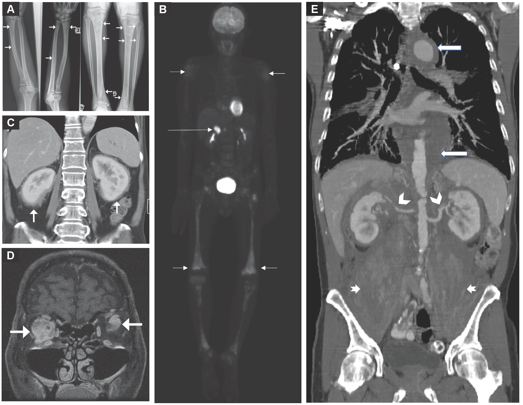
Fig. 1. Imaging findings for Erdheim-Chester disease (ECD). (A) Radiographs of the upper and lower limbs show characteristic bilateral symmetrical involvement of the long bones, with heterogenous sclerosis of the diaphyses and metaphyses (arrows). (B) Whole-body fluorodeoxyglucose (FDG) positron emission tomography (PET) scan shows bilateral and symmetrical raised metabolic activity in the long bones of the upper and lower limbs (short arrows). An extraskeletal site of ECD is in the enlarged, FDG-avid right adrenal gland (long arrow). (C) The “hairy kidney” sign. Contrast-enhanced axial computed tomography (CT) scan shows bilateral and symmetrical irregular soft-tissue infiltration (arrows) in the perirenal spaces. (D) Magnetic resonance imaging (MRI) scan showing orbital ECD lesions. Coronal gadolinium-enhanced fat-suppressed T1-weighted MRI scan shows avid homogeneous enhancement of the intraconal lesions. (E) Retroperitoneal, vascular and muscular involvement in ECD. Coronal enhanced CT scan shows periaortic infiltration along the entire length of the aorta (long arrows) creating a “coated aorta” appearance. The abnormal soft tissue is encasing bilateral renal arteries causing irregular luminal narrowing (arrowheads). There is also diffuse infiltration of bilateral psoas muscles (short arrows).
Renal and retroperitoneum. Approximately 70% of ECD patients have urologic or retroperitoneal involvement. Urological symptoms include abdominal pain, lower urinary tract symptoms, chronic renal insufficiency and renovascular hypertension, but these are uncommon at initial presentation.3,4,12
Histiocytic infiltration of bilateral perirenal spaces manifests on CT scan as low-density soft-tissue infiltrates, giving the “hairy kidney” sign (Fig. 1)—a key imaging feature seen in up to 68% of patients. On MRI scan, the infiltrates are isointense to muscle on T1W and T2W sequences and show mild homogenous enhancement.1,5 Extension to the renal sinus and ureters may result in hydroureteronephrosis. Other complications include renal artery stenosis and chronic kidney disease.12
Adrenal involvement is seen in up to 32% of patients. Imaging findings include diffuse symmetrical bilateral thickening and bulky masses.1,5
Imaging guidelines. Given the multisystemic involvement, a wide array of radiological modalities is needed. Baseline imaging work-up consisting of whole-body FDG PET-CT; contrast-enhanced MRI of the brain; cardiac MRI; and CT of the chest, abdomen and pelvis should be done in all patients to identify disease burden including clinically occult lesions. Imaging follow-up with FDG PET-CT once every 3–6 months is recommended after initiation of treatment. Additionally, organ-specific imaging is recommended every 3 months following treatment initiation, followed by imaging once every 6 months once disease stabilises.1,11
REFERENCES
- Haroche J, Charlotte F, Arnaud L, et al. High prevalence of BRAF V600E mutations in Erdheim-Chester disease but not in other non-Langerhans cell histiocytoses. Blood 2012;120:2700-3.
- Diamond EL, Durham BH, Haroche J, et al. Diverse and Targetable Kinase Alterations Drive Histiocytic Neoplasms. Cancer Discov 2016;6:154-65.
- Antunes C, Graça B, Donato P. Thoracic, abdominal and musculoskeletal involvement in Erdheim-Chester disease: CT, MR and PET imaging findings. Insights Imaging 2014;5:473-82.
- Goyal G, Heaney ML, Collin M, et al. Erdheim-Chester disease: consensus recommendations for evaluation, diagnosis, and treatment in the molecular era. Blood 2020;135:1929-45.
- Boyd LC, O’Brien KJ, Ozkaya N, et al. Neurological manifestations of Erdheim-Chester Disease. Ann Clin Transl Neurol 2020;7:497-506.
- Lachenal F, Cotton F, Desmurs-Clavel H, et al. Neurological manifestations and neuroradiological presentation of Erdheim-Chester disease: report of 6 cases and systematic review of the literature. J Neurol 2006;253:1267-77.
- Wang F, Cao X, Niu N, et al. Multisystemic Imaging Findings in Chinese Patients With Erdheim-Chester Disease. AJR Am J Roentgenol 2019;213:1179-86.
- Drier A, Haroche J, Savatovsky J, et al. Cerebral, facial, and orbital involvement in Erdheim-Chester disease: CT and MR imaging findings. Radiology 2010;255:586-94.
- Wittenberg KH, Swensen SJ, Myers JL. Pulmonary involvement with Erdheim-Chester disease: radiographic and CT findings. AJR Am J Roentgenol 2000;174:1327-31.
- Das JP, Xie L, Riedl CC, et al. Cardiothoracic manifestations of Erdheim-Chester disease. Br J Radiol 2019;92:20190473.
- Diamond EL, Dagna L, Hyman DM, et al. Consensus guidelines for the diagnosis and clinical management of Erdheim-Chester disease. Blood 2014;124:483-92.
- Yelfimov DA, Lightner DJ, Tollefson MK. Urologic manifestations of Erdheim-Chester disease. Urology 2014;84:218-21.
