

Dear Editor,
Percutaneous transcatheter aortic valve implantation (TAVI) has become an established therapy in elderly patients with severe aortic valve stenosis (AS) and an “off-label” option in selected patients with severe aortic regurgitation (AR) at high surgical risk.1 There is also increasing use of TAVI to treat degenerated bioprosthetic surgical aortic valve replacement. Coronary artery occlusion (CAO) is an uncommon but life-threatening complication of TAVI, and occurs when the transcatheter heart valve (THV) pushes the native aortic or surgical valve leaflets outward and occludes the coronary ostia. Furthermore, as TAVI is increasingly performed in younger patients, preserving future coronary artery access has become an important consideration. Recently, catheter-based electrosurgical leaflet laceration with techniques, such as bioprosthetic or native aortic scallop intentional laceration to prevent iatrogenic coronary artery obstruction (BASILICA), has been developed to prevent CAO and to preserve coronary access by splaying open the native or surgical leaflet(s) (Figs. 1A and 1B).
Fig. 1. BASILICA illustration and images of electrosurgical leaflet laceration during TAVI.
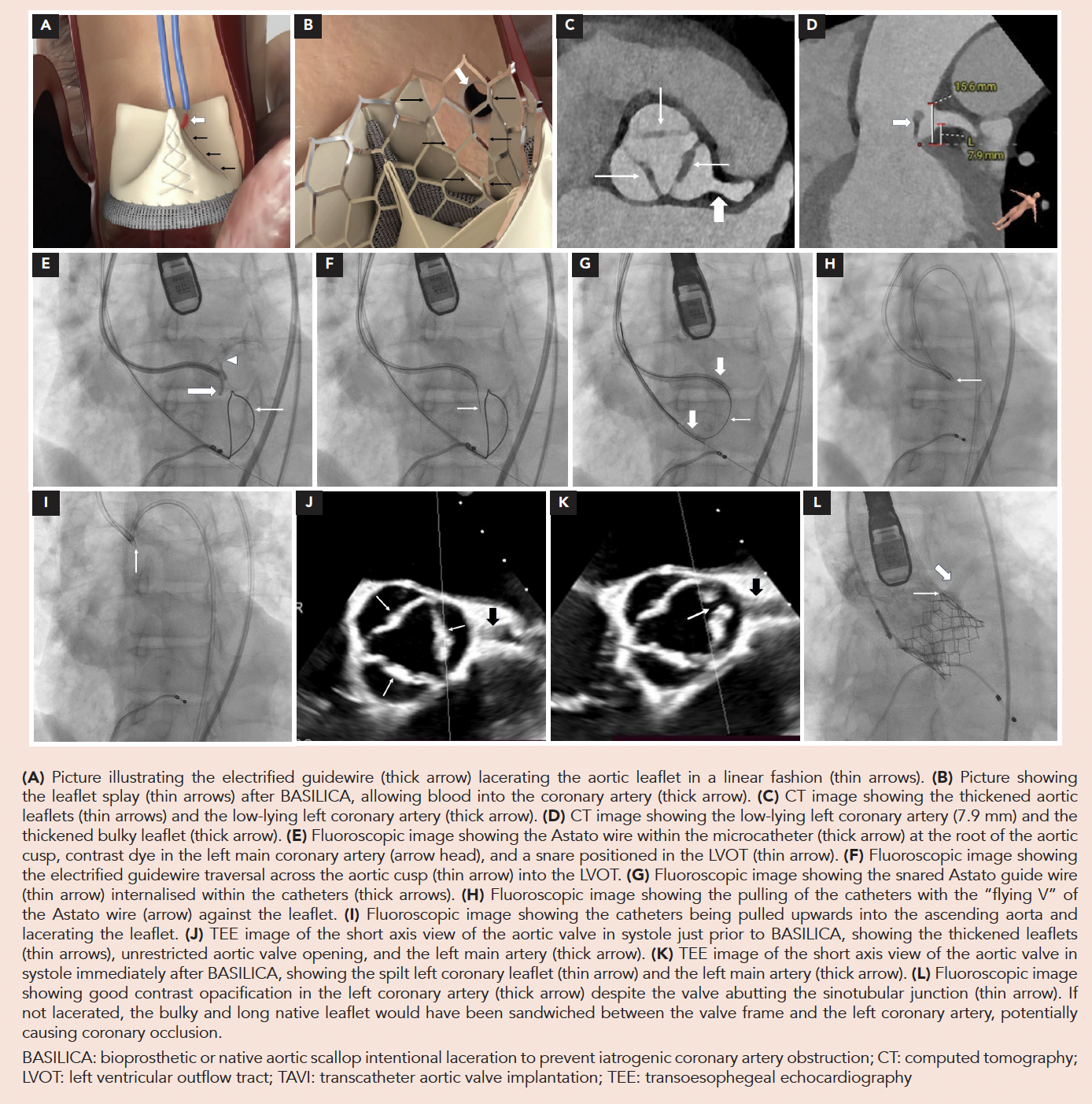
Several studies have demonstrated that in patients undergoing TAVI at elevated risk of CAO, BASILICA is effective in preventing or reducing CAO.2,3 However, widespread use of this technique is limited by procedural complexity. The authors report a case of electrosurgical native leaflet laceration to prevent coronary occlusion during TAVI in a patient with severe AR and at significant risk of CAO.
A 63-year-old male with end-stage renal failure on haemodialysis, and a renal transplant that failed 4 years prior, presented with exertional dyspnoea (New York Heart Association [NYHA] class II). Echocardiography showed normal left ventricular ejection fraction (60%), dilated left ventricle (end-diastole 6.2 cm), and severe AR due to valve malcoaptation. There was no significant AS (aortic valve area by planimetry was 2.2 cm2 by both echocardiography and computed tomography [CT]; mean pressure gradient was mildly elevated at 23 mmHg, consistent with increased forward flow due to the severe AR). Although the patient’s Society of Thoracic Surgeons score of 3.76% for 30-day mortality (22.9% for 30-day mortality/morbidity) indicated moderate surgical risk, he was deemed to be at high surgical risk by the cardiac surgeon owing to frailty, low body weight, previous major gastrointestinal bleeding, and splenic rupture—and was referred for TAVI. CT aortogram showed aortic annulus area 420 mm2 (23 mm diameter), left ventricular outflow tract diameter 25 mm, sinus of Valsalva diameter 29 mm, and aortic leaflets were markedly thickened without calcification (Fig. 1C). The left coronary artery (LCA) take-off height was low at 7.9 mm, and the left coronary leaflet was bulky and long (length 14.1 mm) (Fig. 1D), indicating elevated risk of left CAO. TAVI with BASILICA was planned to reduce the risk of left CAO and to preserve future access to the LCA.
The procedure was performed under general anaesthesia with fluoroscopy and transoesophegeal echocardiography (TEE) guidance. Details of the BASILICA procedure have been described elsewhere.4 In brief, through an 8-French size Amplatz Left 2 curve guide catheter, an Astato 20 (stiff 0.014-inch peripheral angioplasty) wire is positioned at the root of the left aortic cusp below the LCA (Fig. 1E). The wire is electrified using an electrosurgery pen, and advanced through the leaflet into the left ventricular outflow tract (LVOT) (Fig. 1F). A 25 mm Gooseneck snare pre-positioned in the LVOT then snares the Astato wire (Fig. 1G). The Asato wire is pulled back against the leaflet (Fig. 1H), a “flying V” is created and positioned at the leaflet base, and then the “flying V” is electrified and pulled upwards into the ascending aorta to lacerate the leaflet (Fig. 1I). Leaflet laceration was evident by a drop in diastolic pressure and visualisation of split leaflets on TEE (Figs. 1J and 1K). A balloon-expandable 26 mm Sapien 3 Ultra Resilia (Edwards Lifesciences, Irvine, CA, US) THV was successfully implanted (at nominal volume) with upfront cerebral embolic protection (Sentinel system, Boston Scientific, Marlborough, MA, US). Root aortography showed that the LCA was patent (Fig. 1L).
The patient was discharged 2 days post-procedure. At 6 months, he was in NYHA functional class I. Echocardiography revealed satisfactory valve haemodynamics (mean pressure gradient 15 mmHg), no AR, and a reduction in end-diastolic left ventricle size to 5.6 cm.
CAO is a rare complication of TAVI, occurring in <1% of patients; it is however, associated with a high mortality rate of 40% at 30 days.5 Most of such cases are due to direct CAO by the native aortic or surgical leaflets that are pushed outwards by the THV; in a minority of cases, CAO occurs due to sealing of the sinus of Valsalva when the native aortic or surgical leaflets are pushed against the sinotubular junction.
Certain CT measurements have been found to predict an elevated risk of CAO, such as a low coronary height as measured from the aortic annulus (≤10 mm), narrow sinus of Valsalva relative to size of the THV to be implanted with a virtual valve-to-coronary distance of <4 mm, and bulky calcified leaflets (calcium volume >600 mm3).2,6
Electrosurgery leaflet laceration (BASILICA) has been shown to be effective at preventing CAO in patients with native AS or bioprosthetic valve failure.2 Using commercially available equipment, the target leaflet is lacerated, and after THV implantation, the lacerated leaflet is pushed outwards and is splayed open. This triangular space allows coronary blood flow through the THV stent struts. Another benefit of BASILICA is that it will likely facilitate cannulation of the coronary artery, which is relevant in younger patients who may require future percutaneous coronary intervention.7
Data from the BASILICA trials have been promising with rare CAO in patients with the highest risk anatomy. Previously to prevent CAO, a simpler technique—chimney/snorkel stenting, where a stent is implanted at the coronary ostium with protrusion into the aorta—was used. However, there is a lack of data on its longevity with the stent extending beyond the coronary ostium and sandwiched by a THV. There are concerns of inability to re-access the coronary artery, stent deformation, fatal in-stent restenosis or thrombosis, and need for indefinite dual antiplatelet therapy.8.9 In a non-randomised trial comparing BASILICA versus chimney stenting, both therapies were equally effective in preventing CAO; however, there was a numerically higher 1-year cardiovascular mortality with chimney stenting.3 Despite these advantages, there are several limitations of the BASILICA technique. There is a minute residual risk of CAO after BASILICA, particularly if the laceration was not performed from the base of the coronary cusp or directly below the coronary ostium. Hence, coronary protection with a guide catheter and coronary wire may still be necessary. Haemodynamic instability can occur after BASILICA leaflet laceration due to increased AR, although this fortunately, has been rare. As BASILICA is a technically complex procedure, a simpler alternative technique of leaflet modification has been developed—Undermining Iatrogenic Coronary Obstruction with Radiofrequency Needle (UNICORN)—where after leaflet traversal with the electrified Astato wire, the aortic leaflet is dilated with standard angioplasty balloons, and the THV is implanted directly within the aortic leaflet, pushing it away from the coronary ostium.
This case was unique for several reasons. First, this is 1 of the few reported cases of BASILICA in a patient with severe AR worldwide,10 and 1 of the early cases of BASILICA for native aortic valve in Southeast Asia. BASILICA for native aortic valve is a higher risk procedure than BASILICA for degenerated surgical aortic bioprosthesis. Without a radiologically visible sewing ring to help direct the electrified wire, there is a higher risk of aortic root or myocardial injury in native aortic valve BASILICA. Second, the authors demonstrate that TAVI with a balloon-expandable valve is feasible in severe AR with markedly thickened leaflets, despite being an off-label indication. In this case, the thickened leaflets provided adequate surface friction for THV anchoring. This case is different from conventional pure non-calcific severe AR with thin leaflets where the risk of device embolisation or migration is significant using standard TAVI valves. JenaValve, the purpose-designed THV for AR is commercially available (JenaValve Technology, Irvine, CA, US); however, it is not readily available in Southeast Asia. Furthermore, the long-term durability of the JenaValve is unknown, unlike the Sapien 3 Ultra Resilia valve, where the Resilia tissue fixation has been demonstrated, in surgical bioprosthetic valves, to be more durable than previous tissue fixation technology.11 This may be beneficial in a young patient on haemodialysis, although the crimping process that THVs undergo before implantation may affect valve durability, regardless of the tissue fixation technology.
In conclusion, this report demonstrates that electrosurgical leaflet laceration during TAVI is a feasible method of reducing the risk of CAO in patients with high-risk anatomy, and may also preserve coronary artery access which is an important consideration, particularly for younger patients who may have greater need for future percutaneous coronary intervention.
REFERENCES
- Praz F, Borger MA, Lanz J, et al. 2025 ESC/EACTS Guidelines for the management of valvular heart disease. Eur Heart J 2025;46:4635-736.
- Khan JM, Greenbaum AB, Babaliaros VC, et al. BASILICA Trial: One-Year Outcomes of Transcatheter Electrosurgical Leaflet Laceration to Prevent TAVR Coronary Obstruction. Circ Cardiovasc Interv 2021;14:e010238.
- Mangieri A, Richter I, Gitto M, et al. Chimney Stenting vs BASILICA for Prevention of Acute Coronary Obstruction During Transcatheter Aortic Valve Replacement. JACC Cardiovasc Interv 2024;17:742-52.
- Khan JM, Dvir D, Greenbaum AB, et al. Transcatheter Laceration of Aortic Leaflets to Prevent Coronary Obstruction During Transcatheter Aortic Valve Replacement. JACC Cardiovasc Interv 2018;11:677-89.
- Ribeiro HB, Webb JG, Makkar RR, et al. Predictive factors, management, and clinical outcomes of coronary obstruction following transcatheter aortic valve implantation: insights from a large multicenter registry. J Am Coll Cardiol 2013;62:1552-62.
- Khan JM, Kamioka N, Lisko JC, et al. Coronary Obstruction From TAVR in Native Aortic Stenosis: Development and Validation of Multivariate Prediction Model. JACC Cardiovasc Interv 2023;16:415-25.
- Rogers T, Lupu L. Role of leaflet modification in TAVI: to prevent coronary obstruction only or potentially a routine practice? EuroIntervention 2024;20:e733-4.
- Khan JM, Dvir D, Greenbaum AB, et al. Transcatheter Laceration of Aortic Leaflets to Prevent Coronary Obstruction During Transcatheter Aortic Valve Replacement: Concept to First-in-Human. JACC Cardiovasc Interv 2018;11:677-89.
- Lederman RJ, Babaliaros VC, Rogers T, et al. Preventing Coronary Obstruction During Transcatheter Aortic Valve Replacement: From Computed Tomography to BASILICA. JACC Cardiovasc Interv 2019;12:1197-216.
- Kamioka N, Lederman RJ, Khan JM, et al. BI-SILICA During Transcatheter Aortic Valve Replacement for Noncalcific Aortic Insufficiency. JACC Cardiovasc Interv 2018;11:2237-9.
- Kaneko T, Bavaria JE, Thourani VH, et al. Propensity-adjusted 8-year outcomes following bioprosthetic aortic valve replacement: The influence of novel anticalcification technology. JTCVS Open 2026;29:101557.
Written informed consent was obtained for anonymised patient information to be published in this article.
Dr Paul Chiam serves as a proctor for Edwards Lifesciences. The other authors declare they have no affiliations or financial involvement with any commercial organisation with a direct financial interest in the subject or materials discussed in the manuscript.
Dr Paul Toon Lim Chiam, The Heart and Vascular Centre, Mount Elizabeth Medical Centre, 3 Mount Elizabeth, #08-06, Singapore 228510. Email: [email protected]
