

ABSTRACT
Introduction: This study aimed to investigate the relationship between plasma desialylated low-density lipoprotein (desLDL) levels and the severity of coronary artery disease (CAD), and to evaluate the potential value of desLDL as a biomarker for disease stratification and risk prediction.
Methods: This study was conducted at Affiliated Nanhua Hospital, University of South China between December 2024 and June 2025. A total of 162 patients undergoing coronary angiography were enrolled and categorised into a CAD group (n=78) and a non-CAD group (n=84) based on the presence of ≥50% coronary stenosis. Plasma desLDL levels were quantified by liquid chromatography-mass spectrometry (LC-MS). Receiver operating characteristic (ROC) analysis compared the diagnostic performance of desLDL with conventional lipid parameters, and multivariable logistic regression was applied to assess the independent predictive value of desLDL.
Results: Plasma desLDL levels were significantly higher in the CAD group than in controls. Among CAD patients, those with >2 diseased vessels had higher desLDL levels than those with ≤2 vessels, and both exceeded control values (P <0.001). The ROC area under the curve was 0.836 (95% confidence interval [CI] 0.779–0.893); a threshold of 2.25 mmol/L provided 75.6% sensitivity and 84.2% specificity, outperforming LDL cholesterol and total cholesterol. Multivariable analysis identified desLDL as the strongest independent predictor of CAD (adjusted odds ratio 5.85, 95% CI 2.69–12.69). DesLDL elevations were similar in acute myocardial infarction and stable CAD presentations, while both remained significantly higher than controls.
Conclusion: Elevated plasma desLDL levels were significantly associated with the presence of CAD and reflected anatomical disease burden, demonstrating superior predictive performance compared with conventional lipid markers.
CLINICAL IMPACT
What is New
- DesLDL levels were significantly elevated in patients with CAD.
- DesLDL demonstrated superior diagnostic performance compared to traditional lipid parameters for CAD detection.
Clinical Implications
- DesLDL may serve as a novel and independent biomarker for early CAD detection and improved risk stratification.
- DesLDL appears more sensitive for identifying disease burden (number of diseased vessels) than for reflecting disease severity as measured by Gensini score or clinical acuity, suggesting its potential role as a screening rather than a staging tool.
Coronary artery disease (CAD) remains one of the leading causes of death and disability worldwide, imposing a substantial burden on global healthcare systems and socioeconomic development. According to epidemiological data, approximately 197 million individuals were affected by CAD globally in 2019, resulting in an estimated 9.1 million deaths and accounting for 16.1% of total deaths that year.1,2
Atherosclerosis constitutes the fundamental pathological process underlying CAD; it is characterised by lipid accumulation within the arterial intima, endothelial dysfunction and concerted inflammatory cascades that culminate in plaque formation.3,4 These plaques progressively narrow or occlude the coronary lumen, diminish myocardial perfusion and oxygen delivery, and may eventually provoke ischaemic injury or infarction.5,6 Atherosclerosis is particularly prevalent in older adults.7 Over the past decades, numerous risk factors, including dyslipidaemia, smoking, hypertension, diabetes mellitus, ageing, male sex, obesity and chronic inflammation, have been firmly linked to CAD.8,9 Concurrently, an expanding panel of atherosclerosis‑related biomarkers has been introduced into clinical practice;10 however, their sensitivity, specificity, and overall predictive accuracy remain suboptimal, underscoring the need to identify and validate more robust, mechanism‑based indicators.11,12
Given the central role of lipid abnormalities in atherogenesis, low-density lipoprotein cholesterol (LDL-C) is routinely used for cardiovascular risk stratification and therapeutic decision‑making.3,13 Nonetheless, LDL-C levels alone are insufficient, as a considerable proportion of cardiovascular events occur in individuals whose LDL‑C is within recommended targets.14 This discrepancy underscores the need to identify more sensitive atherogenic lipid subtypes. Among modified LDL particles, oxidised LDL (oxLDL) is the most extensively investigated and is now recognised as a pivotal driver of atherosclerotic progression.15,16 OxLDL is rapidly internalised by macrophages via scavenger receptors, triggering foam-cell formation and amplifying local inflammation, thereby accelerating plaque development.17,18 However, antioxidant therapies targeting oxLDL have shown limited clinical efficacy in reducing cardiovascular events,19 suggesting that alternative pathogenic pathways and therapeutic targets warrant exploration.
Desialylated low‑density lipoprotein (desLDL) is among the earliest and most pronounced circulating modifications of native LDL.20,21 Neuraminidases (NEU) remove terminal sialic‑acid residues from the LDL surface,22 generating particles with smaller diameter, higher density and altered lipid composition.23 Compared with native LDL, desLDL displays greater affinity for macrophage scavenger and asialoglycoprotein receptors, thereby enhancing cholesterol uptake and foam‑cell formation.24 Clinically, plasma desLDL levels are elevated 1.5‑ to 6‑fold in patients with CAD, constituting up to 60% of total LDL versus 5–10% in healthy controls.21,25 These observations implicate desLDL in atherogenesis and suggest that it may outperform traditional LDL‑C as a risk marker. Nevertheless, its clinical relevance, particularly for grading disease severity, remains poorly defined and warrants systematic evaluation.
This study quantified plasma desLDL levels in patients undergoing coronary angiography and examined their association with CAD severity, as defined by vessel involvement and Gensini score. The authors further compared the diagnostic performance of desLDL with conventional lipid parameters using receiver operating characteristic (ROC) analysis and evaluated its independent predictive value through multivariable logistic regression. The goal was to determine whether desLDL could function as a novel biomarker for the early detection and refined risk stratification of CAD.
METHODS
Study design and patient recruitment
This prospective, cross‑sectional study enrolled 162 consecutive patients who underwent coronary angiography at the Affiliated Nanhua Hospital, University of South China between December 2024 and June 2025. Exclusion criteria included active infection, autoimmune disease, severe hepatic or renal dysfunction, malignancy or other major systemic illness.
CAD was defined as ≥50% diameter stenosis with compatible symptoms or electrocardiographic changes. Acute myocardial infarction (AMI) was diagnosed when cardiac troponin was elevated with clinical or imaging evidence of acute ischaemia. Type 2 diabetes mellitus (T2DM) was diagnosed when fasting plasma glucose was ≥7.0 mmol/L or 2‑hour glucose was ≥11.1 mmol/L. Hypertension was defined as blood pressure ≥140/90 mmHg on 3 separate readings.
Patients were stratified into a CAD group (≥50% luminal stenosis in ≥1 major coronary artery; n=78) and a non‑CAD group (n=84), and were frequency‑matched for age and sex. The non-CAD group comprised patients who underwent coronary angiography for clinical suspicion of CAD but were found to have no significant coronary stenosis (<50% in all major vessels). Their clinical presentation and referral indications were comparable to those of the CAD group, ensuring comparability. Four senior interventional cardiologists performed the procedures, and 2 investigators, blinded to participants’ clinical data, independently reviewed the angiograms. Within the CAD cohort, patients were further categorised as AMI versus non‑AMI and as ≤2‑vessel versus >2‑vessel disease. Lesion severity was quantified using the Gensini score, a well-established angiographic scoring system that comprehensively accounts for the number, location and degree of coronary artery stenoses. Although the Synergy between Percutaneous Coronary Intervention With Taxus and Cardiac Surgery (SYNTAX) score is also widely used, the Gensini score is particularly suitable for assessing the overall atherosclerotic burden in diverse patient populations, including those with multivessel disease, and has been validated in numerous studies for predicting cardiovascular outcomes (Fig. 1).
Fig. 1. STROBE flowchart.
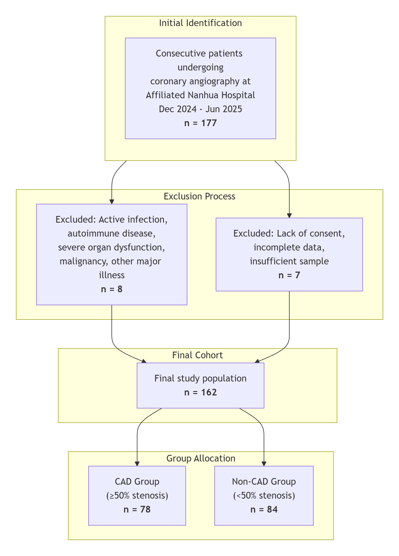 CAD: coronary artery disease; STROBE: strengthening the reporting of observational studies in epidemiology
Clinical and laboratory data
Baseline demographics (age, sex, body mass index [BMI], smoking, hypertension, T2DM) were recorded. Fasting plasma was stored at -80 °C and analysed once. Plasma desLDL was quantified using a targeted liquid chromatography-tandem mass spectrometry (LC-MS/MS) method based on the specific derivatisation of asparagine residues in apolipoprotein B-100, which are exposed upon desialylation. Lipid profile (total cholesterol [TC], LDL‑C, high-density lipoprotein‑cholesterol, triglyceride) was measured on an automated analyser; high-sensitivity C-reactive protein (hs-CRP) was assayed by enzyme-linked immunosorbent assay.
Analytical validation of LC-MS/MS
The analytical performance of the LC-MS/MS method for quantifying desLDL was rigorously validated according to the U.S. Food and Drug Administration guidance for industry on bioanalytical method validation.26 LDL was isolated from 200 µL of plasma using a precipitation-based method with phosphotungstic acid and magnesium chloride. Purity was confirmed by sodium dodecyl sulfate-polyacrylamide gel electrophoresis, showing a single apolipoprotein B-100 (apoB-100) band without significant contamination. The average recovery of spiked desLDL standards was 92.5% ± 3.2%. To prevent ex vivo desialylation, all plasma samples were immediately placed on ice after collection, processed within 2 hours and handled at 4°C throughout. For quantification, a calibration curve was constructed using enzymatically desialylated LDL standards, with a stable isotope-labelled peptide serving as an internal standard to correct for matrix effects and instrument variability.
The method demonstrated excellent performance characteristics. Calibration curves showed strong linearity over the tested range (R²>0.995), with a lower limit of quantification of 0.1 mmol/L. Both intra-assay (coefficient of variation [CV] <8.5%) and inter-assay (CV <10.2%) precision, as well as accuracy (relative error within ±7.0%), were within acceptable limits. Stability studies confirmed that desLDL was stable for at least 6 months at -80°C and through 3 freeze-thaw cycles. To eliminate pre-analytical bias, all clinical samples were randomised and analysed in a single batch by an operator who was blinded to the participants’ CAD status. The final reported concentration of desLDL (in mmol/L) represents the absolute molar concentration of desialylated LDL particles in the plasma sample. This value was calculated from the measured molar concentration of a specific apoB-100-derived peptide, which is exposed and derivatised only in desialylated LDL, under the biochemical assumption that each individual LDL particle contains a single copy of the apoB-100 protein.
Statistical analysis
Analyses were performed in SPSS version 27.0 (IBM Corp, Armonk, NY, US) and R 4.4.1 (R Core Team, Vienna, Austria). Normality was assessed with the D’Agostino-Pearson test. Data were presented as mean ± standard deviation (SD), median (interquartile range [IQR]) or number (%). Between‑group differences were tested with Student’s t‑tests, Mann-Whitney U tests or chi-square tests as appropriate. Spearman correlation assessed the relationship between desLDL and Gensini score. Diagnostic accuracy was evaluated with ROC curves, and areas under the curves (AUCs) were compared with those for LDL‑C and total cholesterol (TC) using DeLong’s test. The optimal desLDL cut‑off was defined by the Youden index. Independent associations with CAD were tested in multivariable logistic regression; covariates with P <0.10 in univariate analysis and variance inflation factor <2.0 were retained. Results were reported as adjusted odds ratios (ORs) with 95 % confidence interval (CIs). A 2‑sided P<0.05 was considered statistically significant, and post‑hoc power analysis confirmed adequate study power.
RESULTS
Baseline characteristics
Compared with individuals without CAD, patients in the CAD group displayed an overall less favourable cardiometabolic profile. BMI was higher in the CAD group (24.34 ± 3.85 versus [vs] 23.04 ± 2.91 kg m-2, P=0.016), and current smoking was more prevalent (47.4% vs 21.4%, P<0.001). Hypertension, T2DM and statin therapy were likewise more common (60.3% vs 32.1%, P<0.001; 33.3% vs 14.3%, P=0.005; 56.4% vs 9.5%, P<0.001, respectively). TC, LDL‑C and lipoprotein(a) were all significantly higher in the CAD group (4.45 ± 0.61 vs 4.27 ± 0.48 mmol/L, P = 0.040; 3.03 ± 1.09 vs 2.51 ± 0.68 mmol/L, P < 0.001; 182.97 ± 77.85 vs 101.87 ± 40.61 mg/L, P<0.001, respectively). Inflammatory burden was also greater in the CAD group. Patients with CAD showed significantly higher hs-CRP (median 2.31 vs 1.09 mg/L) and white blood cell counts (6.95 vs 5.78×10⁹ mg/L, both P<0.001) than the non-CAD group (Table 1).
Table 1. Baseline characteristics of the study participants.
|
Variables |
CAD (n=78) |
Non-CAD (n=84) |
P value |
|
Age, mean ± SD, years |
55.58 ± 11.02 |
55.77 ± 7.97 |
0.895 |
|
Sex: male, no. (%) |
42 (53.8) |
35 (41.7) |
0.156 |
|
BMI, mean ± SD, kg/m2 |
24.34 ± 3.85 |
23.04 ± 2.91 |
0.016 |
|
Smoking, no. (%) |
37 (47.4) |
18 (21.4) |
<0.001 |
|
Hypertension, no. (%) |
47 (60.3) |
27 (32.1) |
<0.001 |
|
T2DM, no. (%) |
26 (33.3) |
12 (14.3) |
0.005 |
|
Statin, no. (%) |
44 (56.4) |
8 (9.5) |
<0.001 |
|
TG, mean ± SD, mmol/L |
1.29 ± 0.56 |
1.20 ± 0.34 |
0.204 |
|
TC, mean ± SD, mmol/L |
4.45 ± 0.61 |
4.27 ± 0.48 |
0.040 |
|
LDL-C, mean ± SD, mmol/L |
3.78 ± 0.74 |
2.82 ± 0.57 |
<0.001 |
|
HDL-C, mean ± SD, mmol/L |
1.09 ± 0.26 |
1.11 ± 0.20 |
0.695 |
|
Lp(a), mean ± SD, mg/L |
182.97 ± 77.85 |
101.87 ± 40.61 |
<0.001 |
|
hs-CRP, median (IQR), mg/L |
2.31 (0.11–8.16) |
1.09 (0.14–1.97) |
<0.001 |
|
WBC, mean ± SD, ×109 mg/L |
6.95 ± 2.14 |
5.78 ± 1.22 |
<0.001 |
BMI: body mass index; CAD: coronary artery disease; HDL-C, high-density lipoprotein cholesterol; hs-CRP, high-sensitivity C-reactive protein; IQR: interquartile range; LDL-C: low-density lipoprotein cholesterol; Lp(a): lipoprotein(a); SD: standard deviation; T2DM: type 2 diabetes mellitus; TC: total cholesterol; TG: triglyceride; WBC: white blood cell count
Differences in plasma desLDL levels between CAD and non-CAD groups
As shown in Fig. 2, plasma desLDL concentrations were significantly elevated in patients with CAD compared with angiographically normal controls (2.64 ± 0.78 vs 1.69 ± 0.58 mmol/L, P<0.001). Within the CAD group, desLDL increased with anatomical burden; individuals harbouring >2 diseased vessels showed higher levels than those with ≤2 vessels (P<0.01), and both subgroups exceeded non‑CAD values (all P<0.001; Fig. 3A). When patients were stratified by Gensini score (≥51 vs <51), there was no statistically significant difference in desLDL levels between the 2 groups (Fig. 3B). In the analysis of clinical presentation, patients with CAD who presented with AMI (n=22) and those without AMI (n=56) exhibited comparable desLDL levels, yet both were markedly higher than those in non‑CAD controls (Fig. 3C). These findings indicate that plasma desLDL is associated with coronary atherosclerotic burden, while its elevation is not necessarily specific to AMI onset.
Fig. 2. Comparison of plasma desLDL levels between CAD and non‑CAD groups. The plasma desLDL concentration was significantly higher in patients with CAD (n=78, 2.64 ± 0.78 mmol/L) than in non-CAD individuals (n=84, 1.69 ± 0.58 mmol/L); Student’s t-test, P<0.001).
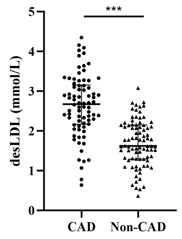

CAD: coronary artery disease; desLDL: desialylated low-density lipoprotein
Fig. 3. Comparison of plasma desLDL levels between CAD subgroups. (A) desLDL levels were significantly higher in CAD patients with ≤2 diseased vessels (n=43) and >2 diseased vessels (n=35) than in non-CAD patients (n=84), with median (IQR) values of 2.50 (2.24–2.84), 2.86 (2.61–3.25) and 1.73 (1.43–1.95) mmol/L, respectively (***P<0.001 for both). Moreover, patients with >2 diseased vessels had significantly higher desLDL levels than those with ≤2 vessels (**P<0.01). (B) desLDL levels were significantly elevated in both CAD patients with Gensini scores <51 (n=39) and ≥51 (n=39) compared to non-CAD controls, with median (IQR) values of 2.73 (2.10–3.07), 2.59 (2.29–3.17) and 1.62 (1.29–2.14) mmol/L, respectively (***P<0.001). No significant difference was observed between the 2 CAD subgroups. (C) desLDL levels were significantly higher in CAD patients without AMI (n=56) and with AMI (n=22) than in non-CAD patients, with median (IQR) values of 2.71 (2.24–3.17), 2.63 (2.11–2.91) and 1.62 (1.29–2.14) mmol/L, respectively (***P<0.001). No significant difference was found between AMI and non-AMI CAD patients.
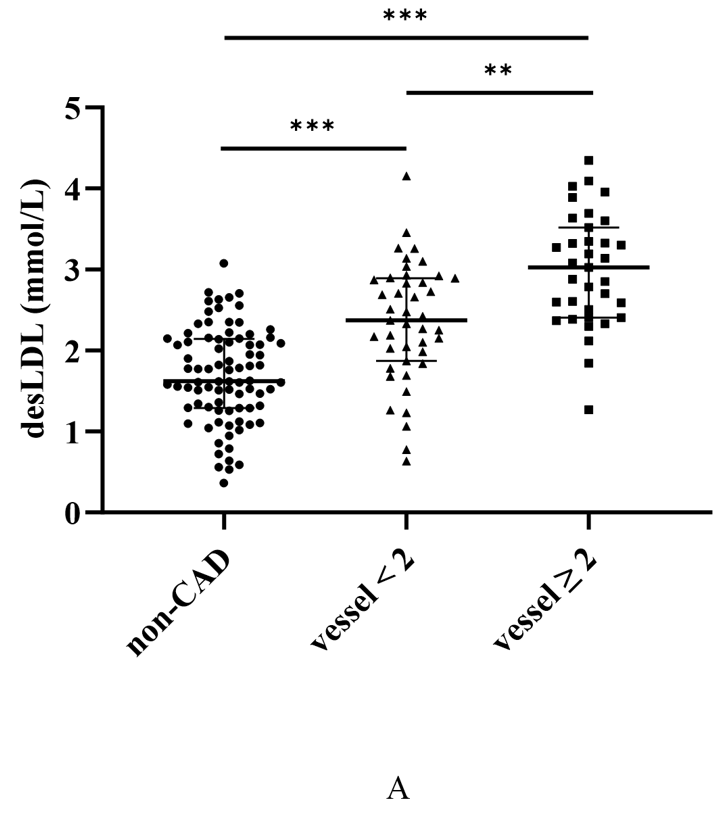
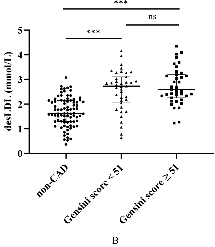
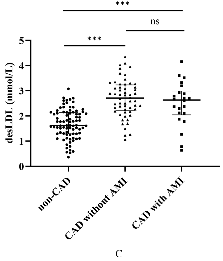
AMI: acute myocardial infarction; CAD: coronary artery disease; desLDL: desialylated low-density lipoprotein; IQR: interquartile range
Fig. 4. ROC curves of desLDL and traditional lipid-related biomarkers for discriminating CAD.
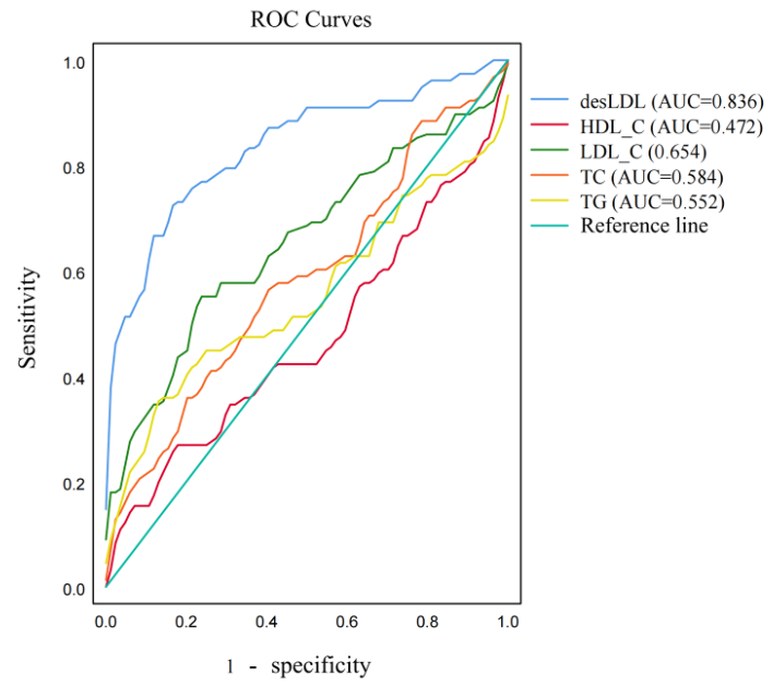
CAD: coronary artery disease; desLDL: desialylated low-density lipoprotein; HDL-C: high-density lipoprotein cholesterol; LDL-C: low-density lipoprotein cholesterol; ROC: receiver operating characteristic; TC: total cholesterol; TG: triglyceride
Diagnostic value of desLDL compared with conventional biomarkers
ROC curve analysis was performed to evaluate the diagnostic accuracy of plasma desLDL for CAD. DesLDL demonstrated excellent discriminative ability with an AUC of 0.836 (95% CI 0.779–0.893). The optimal cut-off value was identified as 2.25 mmol/L, yielding a sensitivity of 75.6% and a specificity of 84.2%.
Comparative analysis revealed that desLDL significantly outperformed all conventional lipid markers. Notably, desLDL’s AUC was substantially higher than that of LDL-C (AUC 0.654, ΔAUC 0.182, P<0.001) and TC (AUC 0.584, ΔAUC 0.252, P<0.001) (Fig. 4).
Association between desLDL and CAD risk
After multivariable adjustment (Table 2), desLDL remained the strongest independent predictor of CAD (OR 5.85, 95 % CI 2.69–12.69, P<0.001). LDL‑C (OR 1.94, 1.13–3.30, P=0.016) and hs‑CRP (OR 1.30, 1.04–1.63, P=0.021) were also significant, as were hypertension (OR 3.57, 1.36–9.35), T2DM (OR 3.98, 1.32–11.99) and smoking (OR 3.58, 1.28–9.99; all P<0.05). BMI was not independently associated with CAD in the adjusted model (OR 1.08, 0.94–1.24, P=0.267).
Table 2. Results of multivariable logistic regression analysis.
|
Variables |
β |
OR (95% CI) |
P value |
|
desLDL |
1.766 |
5.85 (2.69–12.69) |
<0.001 |
|
LDL-C |
0.66 |
1.94 (1.13–3.30) |
0.016 |
|
hs-CRP |
0.263 |
1.30 (1.04–1.63) |
0.021 |
|
BMI |
0.076 |
1.08 (0.94–1.24) |
0.267 |
|
Hypertension |
1.273 |
3.57 (1.36–9.35) |
0.010 |
|
T2DM |
1.381 |
3.98 (1.32–11.99) |
0.014 |
|
Smoking |
1.274 |
3.58 (1.28–9.99) |
0.015 |
BMI: body mass index; CI: confidence interval; desLDL: desialylated low-density lipoprotein; hs-CRP: high-sensitivity C-reactive protein; LDL-C: low-density lipoprotein cholesterol; OR: odds ratio; T2DM: type 2 diabetes mellitus
Multivariable logistic regression analysis was conducted to evaluate the association between selected variables and the presence of coronary artery disease. Results are expressed as ORs with 95% CIs. A P value <0.05 was considered statistically significant.
DISCUSSION
In this study, plasma desLDL was associated with coronary atherosclerotic burden, while its elevation was not necessarily specific to AMI onset. Specifically, plasma desLDL levels rose significantly with increasing angiographic plaque burden. Although a trend towards higher levels was observed in patients with AMI than in those without AMI, this difference did not reach statistical significance. In diagnostic performance, desLDL consistently outperformed conventional lipid measures, including LDL-C and TC, in discriminating CAD (AUC 0.836; sensitivity ≈76%, specificity ≈84%). Importantly, after multivariable adjustment for traditional risk factors, desLDL remained strongly and independently associated with disease presence (adjusted OR ≈5.85). Collectively, these results underscore the premise that lipoprotein quality, specifically the desialylation state, governs coronary risk beyond the quantity of cholesterol alone.
Significant differences in baseline characteristics were observed between patients with CAD and those without. Patients with CAD had a higher BMI and markedly greater prevalence of smoking, hypertension and T2DM, mirroring the established epidemiology of atherosclerotic disease. Smoking and hypertension promote endothelial injury and vascular inflammation, thereby accelerating plaque formation. Patients with CAD also exhibited more pronounced lipid dysregulation and elevated hs-CRP, indicating a more atherogenic systemic milieu. Metabolic disturbances such as T2DM foster small dense LDL and post‑translational LDL modifications, including glycation and desialylation.27,28 Prior work showed that desLDL constitutes ≈35 % of total LDL in diabetes versus ≈12 % in healthy subjects.28 Although the higher T2DM prevalence in this CAD cohort may partly explain the elevated desLDL, the association remained significant after full adjustment, supporting an independent role for desLDL in atherogenesis.
Plasma desLDL was significantly higher in CAD than in controls, consistent with earlier biochemical data reporting a 2.5‑ to 5‑fold reduction in LDL‑bound sialic acid among patients with CAD.29 Subgroup analysis showed that desLDL was higher in patients with >2 diseased vessels than in those with ≤2 diseased vessels. DesLDL was also significantly elevated in both Gensini strata and in both AMI and non-AMI CAD subgroups when each was compared with non-CAD controls.30 However, within‑CAD comparisons based on Gensini category or AMI status did not reach statistical significance, suggesting that desLDL may be more sensitive for detecting disease presence than for quantifying incremental severity.
This pattern echoes that of other glycan‑related biomarkers. Monocyte Siglec‑1 expression is elevated in CAD versus healthy controls but shows no significant differences across CAD phenotypes.31 Likewise, serum total sialic acid is unrelated to lesion extent in stable angina, although it predicts complications in acute coronary syndromes.32,33 The higher desLDL values seen in AMI, together with evidence of dynamic sialic acid upregulation during plaque rupture,33 imply that desialylation may reflect plaque vulnerability and acute inflammatory burden.
Collectively, these data support desLDL as a sensitive and specific marker for the presence of angiographically defined, flow-limiting coronary atherosclerosis (obstructive CAD). These findings suggest that desLDL is elevated even in the context of significant luminal stenosis, highlighting its potential role in identifying patients with established, high-risk atherosclerotic burden. Its ability to distinguish finer gradations of severity may be limited by biological saturation or temporal dynamics; combining desLDL with inflammatory or imaging metrics could therefore enhance the phenotyping of plaque burden and instability.
The superior discriminative power of desLDL over LDL‑C and TC was also confirmed by ROC analysis. Conventional lipids largely quantify particle number, whereas desLDL reflects qualitative particle modification; many CAD patients exhibit normal or target LDL‑C yet harbour structurally altered LDL that confers residual risk.34 These results—and prior data on small dense LDL‑C predicting events despite LDL‑C control35—underscore the value of assessing LDL particle quality in risk stratification. Multivariable modelling further established desLDL as an independent predictor of CAD after controlling for age, sex, LDL‑C, hypertension, T2DM and smoking. Highly desialylated LDL particles may adhere more avidly to the arterial wall and trigger stronger inflammatory responses, fostering plaque growth even in normocholesterolemic individuals. Incorporating desLDL into future risk algorithms could therefore improve identification of high‑risk patients who might benefit from intensified preventive measures.
Mechanistically, NEU1 and NEU3 remove terminal sialic acids from LDL,29 constituting one of the earliest LDL modifications in atherogenesis.36 Loss of negative charge produces smaller, denser, aggregation‑prone particles,34 which evade hepatic LDL receptors but bind macrophage scavenger and asialoglycoprotein receptors with high affinity,29 culminating in foam‑cell formation and plaque expansion.37 In ApoE‑deficient mice, genetic or pharmacological suppression of NEU1/NEU3 markedly attenuates atherosclerosis without altering plasma LDL‑C , confirming desialylation as an LDL‑C-independent driver.
Beyond lipid loading, desLDL intensifies inflammation. Sialic‑acid “self” epitopes normally engage inhibitory sialic acid-binding immunoglobulin-type lectins (Siglecs) on immune cells;38 desialylation disrupts this brake, unleashing macrophage uptake.39 DesLDL‑containing immune complexes activate toll‑like receptors (TLR), inducing cytokine release, macrophage apoptosis and lesion progression.40 NEU1 modulates Siglec-TLR crosstalk, amplifying plaque inflammation.41 These convergent lipid‑accumulation and pro‑inflammatory mechanisms provide a coherent explanation for the strong links between desLDL, CAD presence, and plaque burden observed here.
A pertinent question is how desLDL compares with other established markers of atherogenic particle burden, such as ApoB or LDL-particle number (LDL-P). It is important to recognise that these markers operate on different principles. ApoB and LDL-P quantify the number of atherogenic particles, reflecting the fundamental aetiology of atherosclerosis as a disorder of lipoprotein accumulation. In contrast, this study’s data suggest that desLDL quantifies the functional state, or quality, of these particles. An elevated desLDL level indicates not just the presence of LDL particles, but specifically the presence of a subpopulation that has undergone desialylation—a modification linked to increased atherogenicity and impaired clearance. Therefore, desLDL may provide complementary, rather than redundant, information to ApoB. A patient could theoretically have a normal ApoB level (normal particle number) but a high proportion of desLDL, indicating a high-risk phenotype driven by dysfunctional particles. Conversely, a patient could have a high ApoB but a low desLDL level. Future prospective studies directly comparing these markers head-to-head are needed to define their respective roles. Such research could determine whether desLDL offers incremental prognostic value beyond the simple quantification of particle number provided by ApoB.
Several important limitations merit discussion. First, this single-centre study enrolled only 162 consecutive patients undergoing coronary angiography at a tertiary institution, limiting statistical power and external validity; potential indication bias exists, as patients referred for angiography already had a high pre-test probability of CAD. Second, the cross-sectional design precludes inference of temporality or causality, leaving open the possibility of reverse causation and residual confounding. Third, the authors’ assessment of disease severity relied on the Gensini score; while well-validated, future studies comparing its utility with that of SYNTAX score would be valuable. Fourth, the cohort lacked asymptomatic individuals and detailed baseline medication history, particularly lipid-lowering therapy, preventing adjustment for potential confounding effects and limiting the assessment of desLDL’s role independent of therapeutic interventions. While this study demonstrates a strong association between desLDL and CAD, future research with detailed medication data is required to establish a strong association between desLDL and CAD, future research with detailed medication data is required to establish causality and to determine the true predictive value of desLDL independent of treatment effects.
CONCLUSION
In conclusion, plasma desialylated LDL was significantly elevated in CAD and closely reflected disease burden (number of diseased vessels), outperforming traditional lipid indices. Our findings support desLDL as a novel, independent biomarker for detecting the presence of coronary atherosclerosis rather than for grading its comprehensive severity, although further longitudinal studies are needed to confirm its prognostic value.
Data availability
The datasets generated and/or analysed during the present study are not publicly available but are available from the corresponding author Zhuo Zeng on reasonable request.
REFERENCES
- Wang K, Shi X, Zhu Z, et al. Mendelian randomization analysis of 37 clinical factors and coronary artery disease in East Asian and European populations. Genome Med 2022;14:63.
- Wang H, Abbas KM, Abbasifard M, et al. Global age-sex-specific fertility, mortality, healthy life expectancy (HALE), and population estimates in 204 countries and territories, 1950–2019: a comprehensive demographic analysis for the Global Burden of Disease Study 2019. Lancet 2020;396:1160-203.
- Falk E. Pathogenesis of Atherosclerosis. J Am Coll Cardiol 2006;47:C7-12.
- Zhu Y, Xian X, Wang Z, et al. Research Progress on the Relationship between Atherosclerosis and Inflammation. Biomolecules 2018;8:80.
- Silventoinen K, Lahtinen H, Davey Smith G, et al. Height, social position and coronary heart disease incidence: the contribution of genetic and environmental factors. J Epidemiol Community Health 2023;77:384-90.
- Jiesisibieke ZL, Panter J, Wang M, et al. Mode of transport, genetic susceptibility, and incidence of coronary heart disease. Int J Behav Nutr Phys Act 2023;20:79.
- Kaplan H, Thompson RC, Trumble BC, et al. Coronary atherosclerosis in indigenous South American Tsimane: a cross-sectional cohort study. Lancet 2017;389:1730-9.
- Lechner K, von Schacky C, McKenzie AL, et al. Lifestyle factors and high-risk atherosclerosis: Pathways and mechanisms beyond traditional risk factors. Eur J Prev Cardiol 2020;27:394-406.
- Fularski P, Czarnik W, Dąbek B, et al. Broader Perspective on Atherosclerosis—Selected Risk Factors, Biomarkers, and Therapeutic Approach. Int J Mol Sci 2024;25:5212.
- Tibaut M, Caprnda M, Kubatka P, et al. Markers of Atherosclerosis: Part 1 – Serological Markers. Heart Lung Circ 2019;28:667-77.
- Mietus-Snyder M, Perak AM, Cheng S, et al. Next Generation, Modifiable Cardiometabolic Biomarkers: Mitochondrial Adaptation and Metabolic Resilience: A Scientific Statement From the American Heart Association. Circulation 2023;148:1827-45.
- Annink ME, Kraaijenhof JM, Beverloo CYY, et al. Estimating inflammatory risk in atherosclerotic cardiovascular disease: plaque over plasma? Eur Heart J Cardiovasc Imaging 2025;26:444-60.
- Lewington S, Whitlock G, Clarke R, et al. Blood cholesterol and vascular mortality by age, sex, and blood pressure: a meta-analysis of individual data from 61 prospective studies with 55,000 vascular deaths. Lancet 2007; 3706:1829-39.
- Hamilton P. Evolocumab and clinical outcomes in patients with cardiovascular disease. Ann Clin Biochem 2017;54:511.
- Itabe H, Obama T, Kato R. The Dynamics of Oxidized LDL during Atherogenesis. J Lipids 2011;2011:1-9.
- Di Pietro N, Formoso G, Pandolfi A. Physiology and pathophysiology of oxLDL uptake by vascular wall cells in atherosclerosis. Vasc Pharmacol 2016, 84:1-7.
- Hofmann A, Brunssen C, Morawietz H. Contribution of lectin-like oxidized low-density lipoprotein receptor-1 and LOX-1 modulating compounds to vascular diseases. Vasc Pharmacol 2018;107:1-11.
- Binder CJ, Papac-Milicevic N, Witztum JL. Innate sensing of oxidation-specific epitopes in health and disease. Nat Rev Immunol 2016;16:485-97.
- Jenkins DJA, Kitts D, Giovannucci EL, et al. Selenium, antioxidants, cardiovascular disease, and all-cause mortality: a systematic review and meta-analysis of randomized controlled trials. Am J Clin Nutr 2020;112:1642-52.
- Summerhill VI, Grechko AV, Yet S, et al. The Atherogenic Role of Circulating Modified Lipids in Atherosclerosis. Int J Mol Sci 2019;20:3561.
- Orekhov AN, Tertov VV, Mukhin DN, et al. Modification of low density lipoprotein by desialylation causes lipid accumulation in cultured cells: Discovery of desialylated lipoprotein with altered cellular metabolism in the blood of atherosclerotic patients. Biochem Biophys Res Commun 1989;162:206-11.
- Lillehoj EP, Luzina IG, Atamas SP. Mammalian Neuraminidases in Immune-Mediated Diseases: Mucins and Beyond. Front Immunol 2022;13:883079.
- Tertov VV, Kaplun VV, Sobenin IA, et al. Low-density lipoprotein modification occurring in human plasma possible mechanism of in vivo lipoprotein desialylation as a primary step of atherogenic modification. Atherosclerosis 1998;138:183-95.
- Orekhov AN, Tertov VV, Mukhin DN. Desialylated low density lipoprotein–naturally occurring modified lipoprotein with atherogenic potency. Atherosclerosis 1991;86:153-61.
- Ruelland A, Gallou G, Legras B, et al. LDL sialic acid content in patients with coronary artery disease. Clin Chim Acta 1993;221:127-33.
- U.S. Department of Health and Human Services, Food and Drug Administration, Center for Drug Evaluation and Research, Center for Veterinary Medicine. Bioanalytical Method Validation: Guidance for Industry. Silver Spring, MD: U.S. Food and Drug Administration; 2018 May.
- Stahel P, Xiao C, Hegele RA, et al. The Atherogenic Dyslipidemia Complex and Novel Approaches to Cardiovascular Disease Prevention in Diabetes. Can J Cardiol 2018;34:595-604.
- Sobenin IA, Tertov VV, Orekhov AN. Atherogenic modified LDL in diabetes. Diabetes 1996;45:S35-9.
- Glanz V, Bezsonov EE, Soldatov V, et al. Thirty-Five-Year History of Desialylated Lipoproteins Discovered by Vladimir Tertov. Biomedicines 2022;10:1174.
- Chappey B, Beyssen B, Foos E, et al. Sialic acid content of LDL in coronary artery disease: no evidence of desialylation in subjects with coronary stenosis and increased levels in subjects with extensive atherosclerosis and acute myocardial infarction: relation between desialylation and in vitro peroxidation. Arterioscler Thromb Vasc Biol 1998;18:876-83.
- Xiong Y, Zhou Y, Rong G, et al. Siglec-1 on monocytes is a potential risk marker for monitoring disease severity in coronary artery disease. Clin Biochem 2009;42:1057-63.
- Salomone OA, Crook JR, Hossein-Nia M, et al. Serum sialic acid concentration is not associated with the extent or severity of coronary artery disease in patients with stable angina pectoris. Am Heart J 1998;136:620-3.
- Li M, Qian S, Yao Z, et al. Correlation of serum N-Acetylneuraminic acid with the risk and prognosis of acute coronary syndrome: a prospective cohort study. BMC Cardiovasc Disord 2020;20:404.
- Poznyak AV, Sukhorukov VN, Surkova R, et al. Glycation of LDL: AGEs, impact on lipoprotein function, and involvement in atherosclerosis. Front Cardiovasc Med 2023;10:1094188.
- Zhang J, He L. Relationship between small dense low density lipoprotein and cardiovascular events in patients with acute coronary syndrome undergoing percutaneous coronary intervention. BMC Cardiovasc Disord 2021;21:169.
- Malekmohammad K, Bezsonov EE, Rafieian-Kopaei M. Role of Lipid Accumulation and Inflammation in Atherosclerosis: Focus on Molecular and Cellular Mechanisms. Front Cardiovasc Med 2021;8:707529.
- Faraj TA, Edroos G, Erridge C. Toll-like receptor stimulants in processed meats promote lipid accumulation in macrophages and atherosclerosis in Apoe(-/-) mice. Food Chem Toxicol 2024;186:114539.
- Vladimir IA, Vasily NS, Vasily PK, et al. Chemical composition of circulating native and desialylated low density lipoprotein: what is the difference? Vessel Plus 2017;1:107-15.
- Podrez EA, Febbraio M, Sheibani N, et al. Macrophage scavenger receptor CD36 is the major receptor for LDL modified by monocyte-generated reactive nitrogen species. J Clin Invest 2000;105:1095-108.
- Orekhov AN, Ivanova EA, Melnichenko AA, et al. Circulating desialylated low density lipoprotein. Cor Vasa 2017;59:e149-56.
- Heimerl M, Gausepohl T, Mueller JH, et al. Neuraminidases-Key Players in the Inflammatory Response after Pathophysiological Cardiac Stress and Potential New Therapeutic Targets in Cardiac Disease. Biology (Basel) 2022;11:1229.
The study complied with the Declaration of Helsinki and was approved by the Affiliated Nanhua Hospital, University of South China Ethics Committee (approval number: 2024-ky-173). Written informed consent was obtained from all participants.
The authors declare there are no affiliations with or involvement in any organisation or entity with any financial interest in the subject matter or materials discussed in this manuscript. This study was supported by the Medical and Health Joint Project of Hunan Provincial Natural Science Foundation (No. 2025JJ81098).
Dr Zhuo Zeng, Department of Cardiovascular Medicine, The Fourth Hospital of Changsha, No. 70 Lushan South Road, Yuelu District, Changsha, Hunan, China. Email: [email protected]
