

ABSTRACT
Introduction: Chronic exposure to glucocorticoids (GC) is a leading cause of secondary osteoporosis and fractures. While the skeletal effects of long-term oral GC are well-established, the bone effects of intermittent high-dose intravenous pulse methylprednisolone therapy remain less clearly defined. This study aimed to evaluate its impact on skeletal health in adults not receiving anti-osteoporosis treatment.
Methods: This meta-analysis was conducted according to PRISMA guidelines and prospectively registered. PubMed, Scopus, Embase, and Google Scholar were searched up to September 2025. Studies included longitudinal cohort and case-control studies with appropriate comparators and randomised controlled trials reporting pre- and post-intravenous methylprednisolone bone outcomes.
Results: Eight studies (n=192, 72% females) were included. Pulse intravenous methylprednisolone did not significantly reduce bone mineral density (BMD) at the lumbar spine (mean difference [MD] 0.01 g/cm2; 95% confidence interval [CI] –0.02 to 0.03), femoral neck (MD –0.01 g/cm2; 95% CI –0.03 to 0.01), or total hip (MD –0.03 g/cm2; 95% CI –0.06 to 0.01) over a mean follow-up period of 5.5 months. Trabecular bone score showed no significant pooled change (MD –0.01 g/cm2). In contrast, procollagen type-1 N-terminal propeptide (MD –36.95 ng/mL; 95% CI –56.86 to –17.03) and C-terminal telopeptide (MD –0.19 ng/dL; 95% CI –0.32 to –0.07) significantly decreased. Serum 25-hydroxyvitamin D increased modestly, while serum calcium and parathyroid hormone remained unchanged.
Conclusion: Pulse intravenous methylprednisolone does not cause short-term BMD loss, though it is associated with measurable suppression of bone turnover markers. Larger controlled studies with standardised sampling, peripheral site assessment, and fracture outcomes are needed to determine long-term skeletal risk from pulse regimens.
CLINICAL IMPACT
What is New
- Pulse intravenous methylprednisolone was not associated with short-term loss of lumbar spine or hip bone mineral density (BMD).
- Bone turnover markers were suppressed, indicating early effects on bone remodelling that are not captured by BMD alone.
- Limited fracture reporting precludes firm conclusions regarding fracture risk.
Clinical Implications
- Stable short-term BMD should not be equated with skeletal safety during pulse intravenous methylprednisolone therapy.
• Patients at high fracture risk or receiving repeated/high cumulative doses may still warrant careful skeletal risk assessment and preventive management.
Glucocorticoids (GC) are potent anti-inflammatory agents used for the treatment of various acute autoimmune and inflammatory disorders. Current studies estimate that the prevalence of GC use among countries varies between 0.5 and 21%.1-4 Although they are highly effective, GCs are known to have negative impacts on skeletal health, particularly when administered at high doses and/or for a prolonged period.5-7 Among individuals receiving long-term GC therapy, the estimated prevalence of osteoporosis or fractures is up to 50%, rendering GCs the most common form of secondary (iatrogenic) osteoporosis.8,9 Thus, it is recommended to carry out a fracture risk assessment at GC initiation and to start bone-protective strategies in patients at moderate to high risk of fracture, particularly for those receiving the equivalent of ≥2.5 to 5 mg prednisolone daily for ≥3 months.10
GC-induced skeletal harm is most pronounced in the trabecular-rich regions, such as the vertebrae and proximal femur.11 GC therapy increases the risk of hip and vertebral fractures by at least two- and threefold, respectively, compared with individuals without GC exposure.11 Bone loss occurs most rapidly after GC initiation, with the most rapid decline occurring within the first year of GC therapy at a bone loss rate of 6 to 12%. This later slows to a rate of approximately 3% per year subsequently.12 Consistent with this early bone loss rate, fracture risk is highest within the first 3 to 6 months after GC initiation, reflecting early osteoclast activation, increased osteoblast apoptosis, and reduced osteoblast proliferation.13 In individuals initially commenced on GC, fracture incidence was 5.1% (95% confidence interval [CI] 2.8–8.2%) for vertebral fractures and 2.5% (95% CI 1.2–4.2%) for non-vertebral fractures. For chronic GC users (>6 months), the corresponding incidences were 3.2% (95% CI 1.8–5.0%) and 3.0% (95% CI 0.8–5.9%), respectively.14 More importantly, fracture risk is influenced by current GC dose and cumulative GC exposure.10 Even low doses of GC ≤2.5 mg per day have been associated with increased risk of vertebral fractures,9 and doses above the 2.5 mg per day threshold have been shown to increase the risk of spine and hip fractures.10,15 Cumulative GC exposure is similarly important: higher doses of GCs ≥30 mg per day increase the risk of fragility fractures, with peak fracture incidence at 12 months. Indeed, fracture rate rises with cumulative GC exposure from an average 4.6 per 1000 person-years (95% CI 3.8–5.4) at <0.675 g of cumulative GC to a nearly threefold rate of 13.4 per 1000 person-years (95% CI 10.7–16.7) at a cumulative GC dose of ≥5.4 g.16
Mechanistically, GCs increase skeletal fragility through direct and indirect pathways. GCs directly suppress bone formation by inhibiting osteoblast differentiation, promoting osteoclast apoptosis while reducing osteocyte viability, leading to deterioration of bone microarchitecture and overall quality, ultimately increasing bone fragility. GCs also influence bone remodelling indirectly through suppression of anabolic pathways, including the growth hormone axis, while promoting insulin resistance and muscle catabolism, further compounding skeletal fragility. While the association between chronic GC exposure and adverse skeletal outcomes (including bone loss and fragility fractures) is well established, the impact of intermittent high-dose intravenous pulse methylprednisolone therapy remains less clearly defined.10,17 To date, relatively few studies have directly assessed bone outcomes associated with intermittent pulse methylprednisolone use, and no clear quantitative synthesis of these skeletal effects is available. The authors therefore conducted this systematic review and meta-analysis to clarify the current evidence base. The authors hypothesised that intermittent pulse intravenous methylprednisolone may exert adverse skeletal effects that are detectable in short-term bone turnover markers, even if changes in bone mineral density (BMD) are less readily apparent over short follow-up periods. Accordingly, the authors evaluated the effects of intermittent pulse intravenous methylprednisolone therapy on skeletal health by investigating changes in BMD, bone turnover markers, fracture rate, and bone biochemistry in adults who did not receive osteoporosis therapy.
METHODS
This meta-analysis was conducted in accordance with the Preferred Reporting Items for Systematic Reviews and Meta-Analyses (PRISMA) guidelines.18 As only published data were used, Institutional Review Board approval was not required. The study protocol was prospectively registered in the International Database of Registered Systematic Reviews (https://doi.org/10.17605/OSF.IO/VFMJR).
Selection criteria
Eligible studies were identified through comprehensive searches of PubMed, Scopus, Embase, and Google Scholar, including all publications up to September 2025. Studies were included if they met the following criteria: (1) case–control study, cohort study, longitudinal study or randomised controlled trial; (2) reported at least 1 outcome of interest; (3) conducted in humans; (4) available in full text; and (5) English-language published studies. The authors excluded: (1) duplicate or overlapping cohorts; (2) case–control or cross-sectional studies without control groups; (3) case reports, case series, editorials, protocols, opinion pieces, commentaries, abstracts, and review articles; or (4) grey literature, including conference abstracts and unpublished reports (these sources lacked sufficient methodological detail and complete outcome data for reliable extraction and quality assessment).
Search strategy
The search strategy was developed using the Population, Intervention, Comparison, Outcome (also known as PICO) framework.19 The population group consisted of patients receiving pulse intravenous methylprednisolone, with or without maintenance GC therapy. The intervention was exposure to pulse intravenous methylprednisolone, and the comparator group was baseline (pre-exposure) measurements obtained prior to treatment initiation. Outcomes of interest included BMD, bone turnover markers, fracture incidence, and use of osteoporosis treatment. The full search terms are provided in Supplementary Table S1.
Data extraction
Data extraction was performed independently by 4 investigators using a 3-stage process. In the first stage, titles and abstracts were screened, with full texts retrieved when necessary. In the second stage, full-text articles and their reference lists were reviewed for eligibility. In the third stage, study objectives, methods, and suitability for inclusion were critically appraised. Discrepancies were resolved by consensus, with adjudication by third and fourth reviewers. When essential data were unavailable, corresponding study authors were contacted for clarification or provision of unpublished data.
Extracted data included study characteristics and reported outcomes: patient age; underlying disease conditions resulting in GC therapy; pulse intravenous methylprednisolone regimens; radiological measures (BMD, trabecular bone score [TBS]); bone turnover markers, e.g. procollagen type 1 N-terminal propeptide (P1NP), C-terminal telopeptide (CTx), osteocalcin, hydroxyproline, bone-specific alkaline phosphatase (bsALP); bone biochemistry (serum calcium, 25-hydroxyvitamin D, intact parathyroid hormone; and urine calcium/creatinine ratio); and fracture rate. All included full-text articles were reviewed and approved by all authors prior to final data extraction.
Data synthesis and analysis
Meta-analysis was performed when at least 2 or more studies reported the same outcome in a numerical outcome (e.g. BMD, bone turnover markers, fracture rates). Small study effects and publication bias were assessed using funnel plots (log odds against inverse standard error).
Risk-of-bias assessment
Risk of Bias was evaluated using the Risk of Bias In Non-randomized Studies—of Exposures (ROBINS-E) tool (Supplementary Table S2).20 ROBINS-E evaluates the quality of the study based on 7 domains, including exposure classification, deviations from intended exposures, missing data, outcome measurement, and selective reporting. Each domain was rated as low risk, some concerns, high risk, or very high risk of bias. Four reviewers independently performed assessments for risk of bias, with disagreements resolved through discussion.
Statistical analysis
All analyses were performed using RevMan version 5.3 (Nordic Cochrane Centre, The Cochrane Collaboration, Copenhagen) using random-effects models. When standard deviations (SDs) were not reported, they were estimated from P values or imputed using pooled SDs from comparable studies: √(∑(N × SD²) / ∑N).
For continuous variables, mean difference (MD) was used given its clinical interpretability. Analyses were repeated using standardised mean difference to assess robustness across heterogeneous measurement approaches (e.g. different densitometry platforms and laboratory assays). Apart from statistical significance (P<0.05), a 4% change in BMD was prespecified as the least significant change (LSC).21,22
Statistical heterogeneity was assessed using the I² statistic, with P<0.10 considered as statistically significant, and I² ≥50% indicating substantial heterogeneity. Potential sources of heterogeneity were explored through sensitivity analyses after exclusion of outlier studies (defined a priori as those with opposing effect directions, disproportionately large effect sizes, or high risk of bias), followed by recalculation of pooled effect estimates.
Quality of evidence was assessed by the Grading of Recommendations, Assessment, Development, and Evaluations (also known as GRADE) approach.23
RESULTS
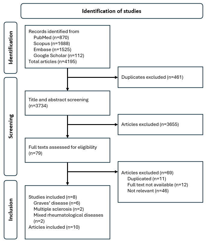
The initial database search identified 4195 records. After removing 461 duplicates, 3734 articles were available for screening. Of these, 3655 articles were excluded based on the predefined eligibility criteria; 79 articles underwent full-text review, of which 69 were excluded, leaving 10 publications (8 studies) in the final review. The 8 included studies comprised 6 cohort studies, 1 retrospective study, and 1 randomised trial. Of the 8 studies, 4 studies recruited patients with Graves’ disease, 2 studies included patients with multiple sclerosis, and 2 studies recruited individuals with mixed rheumatological conditions (Fig. 1). Table 1 summarises the study characteristics and sample sizes ranging from 10 to 45 participants, yielding a total sample of 192 patients (72% female). Means (SDs) of age and body mass index were 49.4 (18.13) years and 25.2 (8.56) kg/m2, respectively.
Table 1. Summary of included studies.
Fig. 1. PRISMA flow chart.
Bone mineral density
Eight studies (10 publications)24-33 reported BMD outcomes before and after pulse methylprednisolone. One study34 reported T-scores and Z-scores only, which could not be reliably converted to BMD values; hence, it was excluded from the analysis. No significant changes were observed in BMD at femoral neck (MD -0.01 g/cm²; 95% CI -0.03 to 0.01; quality of evidence: low ⊕⊕OO), the lumbar spine (MD 0.01 g/cm²; 95% CI -0.02 to 0.03; quality of evidence: low ⊕⊕OO), or total hip (MD -0.03 g/cm²; 95% CI -0.06 to 0.01; quality of evidence: low ⊕⊕OO) following pulse methylprednisolone therapy (all I² = 0%; fixed-effect models applied) that was administered over a weighted mean of 5.5 months (range 3 to 12 months). The lower limit of 95% CI for the BMD changes at femoral neck and lumbar spine did not exceed the prespecified LSC. Of the 2 studies24,28 that reported TBS, no significant difference was detected (MD -0.02; 95% CI -0.10 to 0.07; I² = 0%; fixed-effect; quality of evidence: very low ⊕OOO) (Fig. 2).
Fig. 2. Forest plots of bone mineral density and trabecular bone scores.
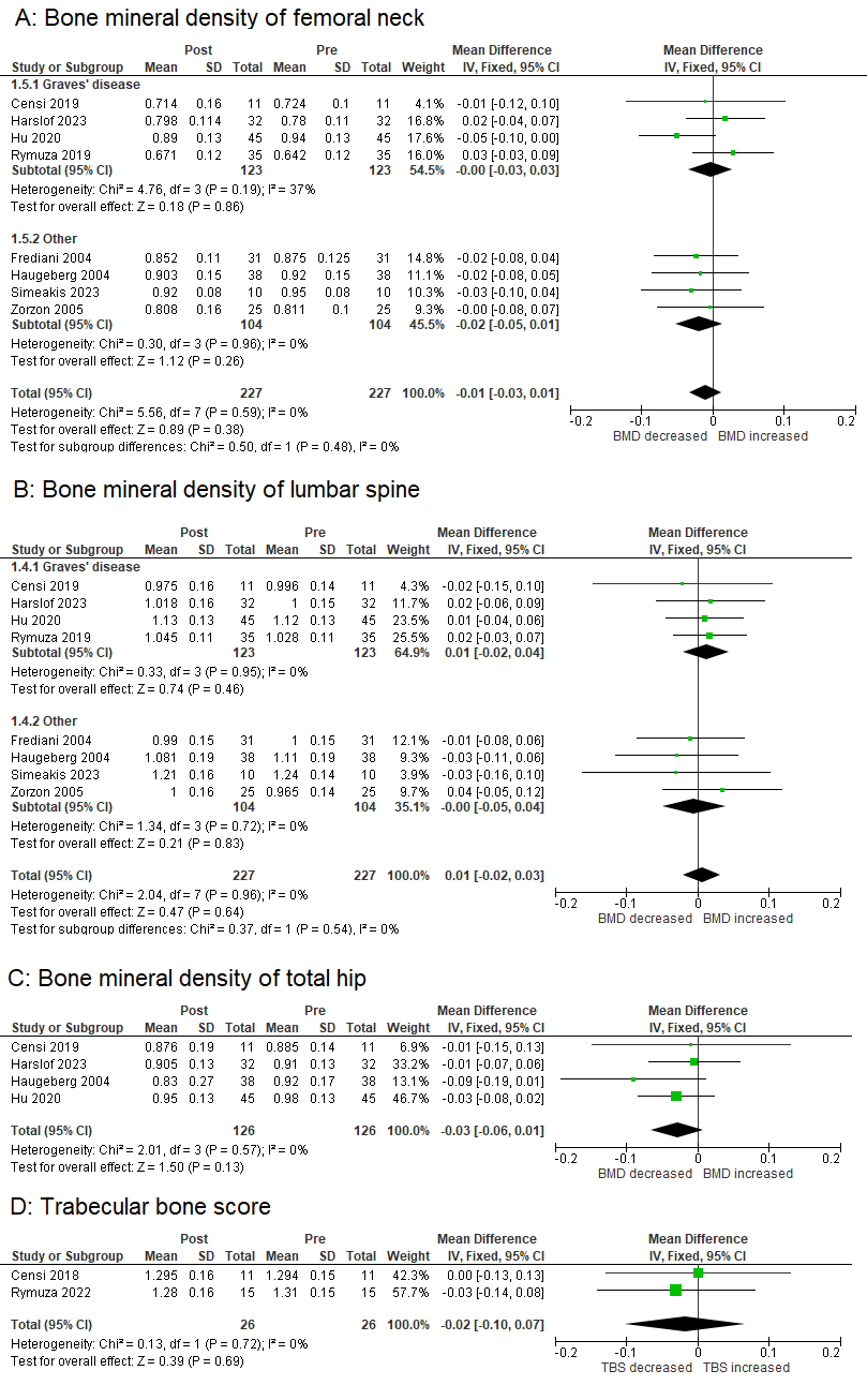
BMD: bone mineral density; CI: confidence interval; df: degrees of freedom; IV: inverse variance; SD: standard deviation; TBS: trabecular bone score
Bone turnover markers
Five studies24,25,27,30,32 reported bone turnover markers. Both P1NP (MD -36.95 ng/mL; 95% CI -56.86 to -17.03; quality of evidence: low ⊕⊕OO) and CTx (MD -0.19 ng/dL; 95% CI -0.32 to -0.07; quality of evidence: low ⊕⊕OO) showed significant reductions following pulse methylprednisolone treatment (I²≥50%; random-effects models applied) (Fig. 3).
Fig. 3. Forest plots of bone turnover markers.
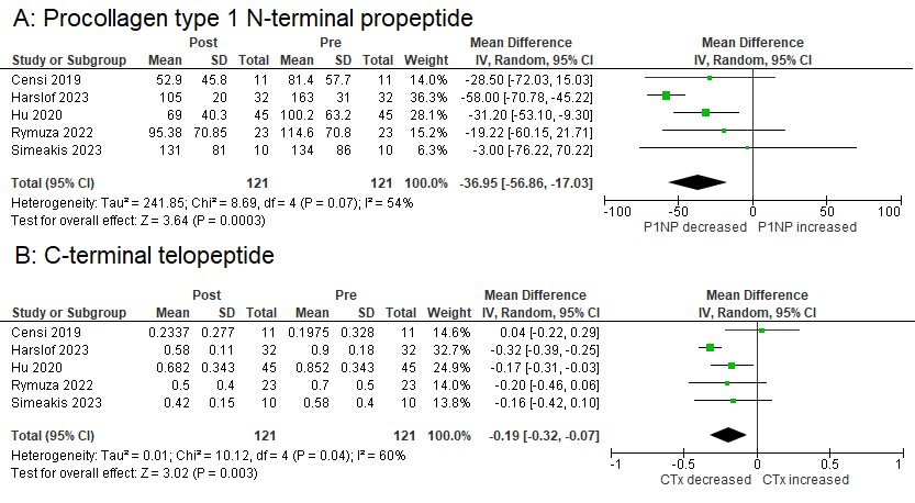
CI: confidence interval; CTx: C-terminal telopeptide; df: degrees of freedom; IV: inverse variance; P1NP: procollagen type 1 N-terminal propeptide; SD: standard deviation
Biochemistry
Serum 25-hydroxyvitamin D levels significantly increased after treatment (MD 1.03 ng/mL; 95% CI 0.24 to 1.83; I² = 0%; fixed-effect; quality of evidence: low ⊕⊕OO).24,27,29,30,32 No significant changes were observed in serum calcium29,30,32 or intact parathyroid hormone24,27,29,30,32 concentrations (Fig. 4).
Fig. 4. Forest plots of bone biochemistry.
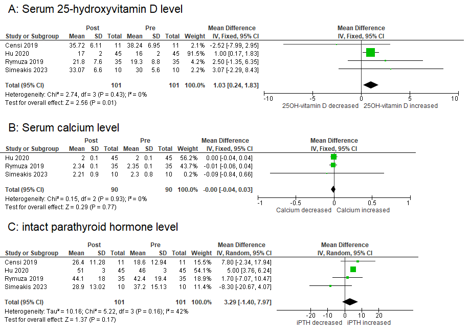
CI: confidence interval; df: degrees of freedom; iPTH: intact parathyroid hormone; IV: inverse variance; SD: standard deviation
Subgroup analysis
A subgroup analysis comparing Graves’ disease24,25,27-29,32 with other conditions (multiple sclerosis30,31 and rheumatological diseases) revealed no significant changes in BMD within either subgroup. Both P1NP and CTx significantly decreased in the Graves’ disease subgroup. In contrast, only a single study was available in the non-Graves’ disease subgroup, and no significant changes were detected (Fig. 2).
Sensitivity analysis
The authors repeated all analyses, including only studies with an overall low risk of bias.25-27,29,30,32 The statistical significance of the results remained unchanged (Table 2).
Table 2. Quality of evidence and sensitivity analysis.
Publication bias
All funnel plots were examined, and no evidence of asymmetry was identified, though the small number of studies may limit interpretability. (Supplementary Fig. S1–9)
DISCUSSION
In this meta-analysis, intermittent pulse methylprednisolone was not associated with a measurable short-term decline in BMD at the lumbar spine, femoral neck or total hip; there was also no consistent change in TBS over a mean follow-up period of 5.5 months. Intravenous pulse methylprednisolone therapy significantly reduced bone turnover markers, despite notable study heterogeneity. The findings suggest that pulse intravenous methylprednisolone can produce early detectable changes in bone remodelling that precede changes in BMD. This contrasts with chronic GC therapy, which is well known to cause rapid changes in BMD within 3 months of GC initiation, predisposing to early fracture risk.35,36
GCs suppress bone formation primarily by inhibiting osteoblast differentiation and activity, in part through inhibition of the Wnt signalling pathway.36,37 GCs also increase the expression of Dickkopf-1 and sclerostin, reduce proliferation of osteoprogenitor cells, and promote differentiation of mesenchymal stem cells into adipocytes rather than osteoblasts.36 These effects result in a sustained reduction in osteogenesis. The low P1NP levels observed in this study are consistent with this biology, and suggest that even intermittent high-dose methylprednisolone exposure may lead to prolonged suppression of osteoblast function lasting for months to years.35,38 The interpretation of reduced CTx is more complex. GCs have mixed and potentially biphasic effects on bone resorption. Initially, GC use has been associated with early bone resorption driven by increased osteoclast activity and attenuation of osteoblast-lineage suppression.38 Over time, osteoclast activity declines due to reduced receptor activator of nuclear factor kappa-B ligand production from suppressed osteoblasts, leading to variable trajectories in resorption markers.38 Apart from the effect of GCs on osteoclasts, GCs can induce osteocyte apoptosis, leading to variable levels of CTx at different time points.39 In pulse methylprednisolone regimens, the timing of sampling is therefore particularly important: CTx measured during or soon after a pulse may be elevated whereas the CTx measured weeks later may be reduced.40 Thus, the concurrent decreases in P1NP and CTx most likely reflect overall suppression of bone remodelling, with a more prominent and sustained inhibitory effect on bone formation. Clinically, this pattern is consistent with a low-turnover skeletal state and may explain the lack of significant BMD changes after a short course of intravenous pulse methylprednisolone therapy. Furthermore, the drug-free intervals in pulse methylprednisolone therapy may allow partial recovery of remodelling signals and normalisation of osteoclast activity.41,42 Nevertheless, persistent suppression of bone formation may still have implications for longer-term bone health.
TBS provides a surrogate index of trabecular bone architecture and predicts fracture risk independent of BMD.43 In this analysis, no significant decline in TBS was observed after pulse intravenous methylprednisolone therapy. However, individual studies have reported divergent results. Rymuza et al.28 reported a decline in TBS in individuals with mild to moderate Graves’ disease orbitopathy treated with pulse intravenous methylprednisolone, whereas earlier studies by Censi et al.24 observed no demonstrable effect of pulse intravenous methylprednisolone on TBS in individuals with Graves’ orbitopathy. Clinically, this suggests that the effect of pulse intravenous methylprednisolone on TBS may be modest and context-specific, and that absence of decline in TBS does not necessarily exclude deterioration in bone quality in susceptible subgroups.
The results are broadly consistent with comparative studies suggesting less BMD loss with pulse regimens compared with chronic daily GCs. Frediani et al.33 conducted a randomised trial of 31 women with active rheumatoid arthritis who received either chronic oral methylprednisolone or intermittent methylprednisolone pulse therapy. After 12 months, chronic methylprednisolone therapy led to significant declines in BMD at the total hip, femur, and lumbar spine while no change in BMD was observed in the pulse methylprednisolone group.33 Handa et al.34 studied 49 patients with dermatological conditions treated with daily prednisolone or intravenous dexamethasone at 4-weekly intervals. The authors found that individuals receiving intermittent intravenous dexamethasone experienced less reduction in lumbar spine T-score than those treated with daily oral prednisolone, consistent with differential effects across trabecular-rich (spine) versus cortical-rich (radius) bone by GC regimen. Among the limited studies reporting fracture outcomes, Zonana-Nacach et al.44 found in 539 patients with systemic lupus erythematosus that higher cumulative prednisone exposure was significantly associated with osteoporotic fractures, whereas each additional intravenous methylprednisolone pulse was associated with only a small, non-significant increase in fracture risk (relative risk 1.3, 95% CI 1.0–1.8; P=0.07). Similarly, in the retrospective 10-year study by Cheng et al.45, no vertebral compression fractures were reported among 46 patients with active systemic sclerosis treated with pulse methylprednisolone therapy, although 21 patients had also received long-term low-dose oral glucocorticoids.
From a clinical standpoint, these findings suggest that short-term stability in BMD following pulse intravenous methylprednisolone should be interpreted cautiously. Baseline fracture risk assessment remains important, especially in patients with high underlying osteoporosis risk or recurrent pulse methylprednisolone exposure. While BMD is unchanged, changes in bone turnover markers are detectable and early fracture risk in GC exposure can be driven by markers not captured in bone imaging. Notably, the absence of a measurable short-term decline in BMD should not be equated with absence of fracture risk, as GC exposure may impair bone strength through mechanisms not fully captured by areal BMD alone.
Furthermore, early fracture risk associated with chronic oral GC therapy could only be partly explained by BMD loss, as excess GC also impairs bone mineralisation and microarchitecture, and could predispose to falls via muscle myopathy. Thus, clinicians should avoid over-reliance on BMD alone and consider vertebral fracture assessment, prior fracture history, fall risk, or even consider using TBS where available.9,35 Most of the studies in this analysis have focused on assessment of bone density in lumbar spine and hip. Handa et al. reported that patients receiving intermittent intravenous dexamethasone had a significantly greater decline in radial head BMD compared with those on daily oral prednisolone, reinforcing the variable sensitivities of skeletal cells to GC regimens.34 Distal radius BMD may be particularly relevant in younger individuals in predicting upper-limb fracture risk,46 and may need to be considered in young individuals receiving intermittent pulse methylprednisolone therapy.47,48 Finally, current guidance and risk prediction tools have not addressed the implications of pulse intravenous methylprednisolone therapy. The Fracture Risk Assessment Tool, also known as FRAX (University of Sheffield, Sheffield, UK), treats GC exposure dichotomously with dose adjustments designed for daily regimens, making translation to intermittent high-dose pulses challenging. Until further evidence emerges, patients undergoing pulse intravenous methylprednisolone treatment with a high risk of fractures should be managed in the same way as those on long-term oral glucocorticoids. This includes providing calcium and vitamin D supplementation, along with standard lifestyle measures that align with the principles for preventing glucocorticoid-induced osteoporosis.
This analysis has several strengths, including a well-structured research question defined within a framework, and formal risk-of-bias assessment that improved methodological rigour and transparency. Nonetheless, several limitations should be acknowledged. Only 8 studies involving 192 participants were included, which restricts statistical power and limits robustness and generalisability of the pooled findings. Moreover, the mean follow-up of approximately 5.5 months may be insufficient to detect clinically meaningful changes in BMD and is inadequate for assessing fracture outcomes, which generally require longer observation periods. Additionally, there was substantial clinical heterogeneity across the included studies. The study populations varied widely, including individuals with Graves’ disease,49,50 multiple sclerosis,51,52 and rheumatological or other inflammatory conditions,53-55 all of which may independently influence bone metabolism through differences in inflammatory burden,56 mobility,57,58 endocrine status,59 nutritional factors,60 and concomitant therapies.61 In addition, pulse intravenous methylprednisolone regimens differed across studies in dose, cumulative exposure, frequency, and duration, which may differentially affect skeletal outcomes. This variability reflects differences in the design and clinical context of the available primary studies and thus represents an inherent limitation of the current evidence base. Furthermore, inconsistent reporting of incident fractures, limited TBS data, and heterogeneity in bone turnover marker assays and sampling time points further constrained interpretation. Although within-person pre-/post-comparisons may reduce some confounding, the small number of studies precluded meaningful subgroup analyses. Accordingly, the findings should be interpreted cautiously, and larger longitudinal studies with longer follow-up are needed.
CONCLUSION
In conclusion, pulse intravenous methylprednisolone therapy does not significantly alter spine or hip BMD over the short-term, but it suppresses bone formation and resorption markers, indicating early effects on bone remodelling. Although pulse regimens may be less detrimental to BMD than chronic daily GC exposure, current evidence is insufficient to conclude that fracture risk is negligible, particularly among high-risk patients or those receiving repeated or high cumulative doses. From a clinical perspective, preservation of short-term BMD should not be equated with skeletal safety; baseline fracture risk assessment remains important, and clinicians should not rely on BMD alone when evaluating patients receiving pulse intravenous methylprednisolone. Future well-designed controlled studies should directly compare pulse and chronic regimens with standardised timing of turnover marker sampling, incorporate bone quality assessments such as TBS at peripheral skeletal sites such as the radius, and include fracture and fall outcomes.
Table S1. Search strategy.
Table S2. Risk of Bias (ROBINS-E).
REFERENCES
- Einarsdottir MJ, Ekman P, Trimpou P, et al. High prescription rate of oral glucocorticoids in children and adults: A retrospective cohort study from Western Sweden. Clin Endocrinol (Oxf) 2020;92:21-8.
- Walsh LJ, Wong CA, Pringle M, Tattersfield AE. Use of oral corticosteroids in the community and the prevention of secondary osteoporosis: a cross sectional study. BMJ 1996;313:344-6.
- Gudbjornsson B, Juliusson UI, Gudjonsson FV. Prevalence of long term steroid treatment and the frequency of decision making to prevent steroid induced osteoporosis in daily clinical practice. Ann Rheum Dis 2002;61:32-6.
- Waljee AK, Rogers MA, Lin P, et al. Short term use of oral corticosteroids and related harms among adults in the United States: population based cohort study. BMJ 2017;357:j1415.
- Souverein PC, Berard A, Van Staa TP, et al. Use of oral glucocorticoids and risk of cardiovascular and cerebrovascular disease in a population based case-control study. Heart 2004;90:859-65.
- Fardet L, Kassar A, Cabane J, et al. Corticosteroid-induced adverse events in adults: frequency, screening and prevention. Drug Saf 2007;30:861-81.
- Broersen LH, Pereira AM, Jorgensen JO, et al. Adrenal Insufficiency in Corticosteroids Use: Systematic Review and Meta-Analysis. J Clin Endocrinol Metab 2015;100:2171-80.
- Compston J. Glucocorticoid-induced osteoporosis: an update. Endocrine 2018;61:7-16.
- Van Staa TP, Leufkens HG, Abenhaim L, et al. Use of oral corticosteroids and risk of fractures. J Bone Miner Res 2000;15:993-1000.
- Humphrey MB, Russell L, Danila MI, et al. 2022 American College of Rheumatology Guideline for the Prevention and Treatment of Glucocorticoid‐Induced Osteoporosis. Arthritis Care & Research 2023;75:2405-19.
- van Staa TP, Leufkens HG, Cooper C. The epidemiology of corticosteroid-induced osteoporosis: a meta-analysis. Osteoporos Int 2002;13:777-87.
- LoCascio V, Bonucci E, Imbimbo B, et al. Bone loss in response to long-term glucocorticoid therapy. Bone Miner 1990;8:39-51.
- Canalis E, Mazziotti G, Giustina A, et al. Glucocorticoid-induced osteoporosis: pathophysiology and therapy. Osteoporos Int 2007;18:1319-28.
- Amiche MA, Albaum JM, Tadrous M, et al. Fracture risk in oral glucocorticoid users: a Bayesian meta-regression leveraging control arms of osteoporosis clinical trials. Osteoporos Int 2016;27:1709-18.
- van Staa TP, Leufkens HG, Abenhaim L, et al. Oral corticosteroids and fracture risk: relationship to daily and cumulative doses. Rheumatology (Oxford) 2000;39:1383-9.
- Balasubramanian A, Wade SW, Adler RA, et al. Glucocorticoid Exposure and Fracture Risk in a Cohort of US Patients With Selected Conditions. J Bone Miner Res 2018;33:1881-8.
- Paccou J, Yavropoulou MP, Naciu AM, et al. Prevention and treatment of glucocorticoid-induced osteoporosis in adults: recommendations from the European Calcified Tissue Society. Eur J Endocrinol 2024;191:G1-17.
- Moher D, Liberati A, Tetzlaff J, et al. Preferred reporting items for systematic reviews and meta-analyses: the PRISMA statement. BMJ 2009;339:b2535.
- Eriksen MB, Frandsen TF. The impact of patient, intervention, comparison, outcome (PICO) as a search strategy tool on literature search quality: a systematic review. J Med Libr Assoc 2018;106:420-31.
- Higgins JPT, Morgan RL, Rooney AA, et al. A tool to assess risk of bias in non-randomized follow-up studies of exposure effects (ROBINS-E). Environ Int 2024;186:108602.
- Eastell R, Rosen CJ, Black DM, et al. Pharmacological Management of Osteoporosis in Postmenopausal Women: An Endocrine Society* Clinical Practice Guideline. J Clin Endocrinol Metab. 2019;104:1595-622.
- Lee CC, Wang CY, Yen HK, et al. Zoledronate Sequential Therapy After Denosumab Discontinuation to Prevent Bone Mineral Density Reduction: A Randomized Clinical Trial. JAMA Netw Open 2024;7:e2443899.
- Schunemann HJ, Brennan S, Akl EA, et al. The development methods of official GRADE articles and requirements for claiming the use of GRADE – A statement by the GRADE guidance group. J Clin Epidemiol 2023;159:79-84.
- Censi S, Manso J, Pandolfo G, et al. Bone turnover markers, BMD and TBS after short-term, high-dose glucocorticoid therapy in patients with Graves’ orbitopathy: a small prospective pilot study. J Endocrinol Invest 2019;42:859-65.
- Harslof T, Hikmet R, Ebbehoj E, et al. Bone turnover decreases and bone structure improves during treatment with weekly high-dose methylprednisolone for 12 weeks in Graves’ orbitopathy. Endocrine 2023;82:664-72.
- Haugeberg G, Griffiths B, Sokoll KB, et al. Bone loss in patients treated with pulses of methylprednisolone is not negligible: a short term prospective observational study. Ann Rheum Dis 2004;63:940-4.
- Rymuza J, Gutowska K, Kurpios-Piec D, et al. Decrease in Bone Formation and Bone Resorption during Intravenous Methylprednisolone Pulse Therapy in Patients with Graves’ Orbitopathy. J Clin Med 2022;11:5005.
- Rymuza J, Pelewicz K, Przedlacki J, et al. Therapy With Intravenous Methylprednisolone Pulses Is Associated With Loss of Bone Microarchitecture in Trabecular Bone Score -Assessment Among Patients With Moderate-to-Severe Graves’ Orbitopathy: A Pilot Study. Front Endocrinol (Lausanne) 2022;13:893600.
- Rymuza J, Popow M, Zurecka Z, et al. Therapy of moderate-to-severe Graves’ orbitopathy with intravenous methylprednisolone pulses is not associated with loss of bone mineral density. Endocrine 2019;64:308-15.
- Simeakis G, Anagnostouli M, Fakas N, et al. High-Dose Intravenous Steroid Treatment Seems to Have No Long-Term Negative Effect on Bone Mineral Density of Young and Newly Diagnosed Multiple Sclerosis Patients: A Pilot Study. Biomedicines. 2023;11:603.
- Zorzon M, Zivadinov R, Locatelli L, et al. Long-term effects of intravenous high dose methylprednisolone pulses on bone mineral density in patients with multiple sclerosis. Eur J Neurol 2005;12:550-6.
- Hu YX, Zheng RD, Fan YF, et al. The effects of bone metabolism in different methylprednisolone pulse treatments for Graves’ ophthalmopathy. Exp Ther Med 2020;19:333-8.
- Frediani B, Falsetti P, Bisogno S, et al. Effects of high dose methylprednisolone pulse therapy on bone mass and biochemical markers of bone metabolism in patients with active rheumatoid arthritis: a 12-month randomized prospective controlled study. J Rheumatol 2004;31:1083-7.
- Handa S, Singh G, Arora AK, et al. Effect of intravenous pulse dexamethasone versus daily oral prednisolone on bone mineral density in dermatology patients: Is it a site-specific response? Indian J Dermatol Venereol Leprol 2018;84:174-8.
- Weinstein RS. Clinical practice. Glucocorticoid-induced bone disease. N Engl J Med 2011;365:62-70.
- Hofbauer LC, Compston JE, Saag KG, et al. Glucocorticoid-induced osteoporosis: novel concepts and clinical implications. Lancet Diabetes Endocrinol. 2025;13:964-79.
- Gado M, Baschant U, Hofbauer LC, et al. Bad to the Bone: The Effects of Therapeutic Glucocorticoids on Osteoblasts and Osteocytes. Front Endocrinol (Lausanne) 2022;13:835720.
- Seibel MJ, Cooper MS, Zhou H. Glucocorticoid-induced osteoporosis: mechanisms, management, and future perspectives. Lancet Diabetes Endocrinol 2013;1:59-70.
- Brabnikova Maresova K, Pavelka K, Stepan JJ. Acute effects of glucocorticoids on serum markers of osteoclasts, osteoblasts, and osteocytes. Calcif Tissue Int 2013;92:354-61.
- Dovio A, Perazzolo L, Osella G, et al. Immediate fall of bone formation and transient increase of bone resorption in the course of high-dose, short-term glucocorticoid therapy in young patients with multiple sclerosis. J Clin Endocrinol Metab 2004;89:4923-8.
- Buckley L, Humphrey MB. Glucocorticoid-Induced Osteoporosis. N Engl J Med 2018;379:2547-56.
- Farraye FA, Melmed GY, Lichtenstein GR, et al. ACG Clinical Guideline Update: Preventive Care in Inflammatory Bowel Disease. Am J Gastroenterol 2025;120:1447-73.
- Leib ES, Winzenrieth R. Bone status in glucocorticoid-treated men and women. Osteoporos Int 2016;27:39-48.
- Zonana-Nacach A, Barr SG, Magder LS, et al. Damage in systemic lupus erythematosus and its association with corticosteroids. Arthritis Rheum 2000;43:1801-8.
- Cheng H, Yu Z, Yan CL, et al. Long-Term Efficacy and Low Adverse Events of Methylprednisolone Pulses Combined to Low-Dose Glucocorticoids for Systemic Sclerosis: A Retrospective Clinical Study of 10 Years’ Follow-Up. J Inflamm Res 2022;15:4421-33.
- Matkovic V, Goel P, Mobley SL, et al. Decreased bone mass in adolescents with bone fragility fracture but not in young children: a case-control study. Front Endocrinol (Lausanne) 2023;14:1124896.
- Cauley JA, Lui LY, LeBoff MS, et al. New Challenges: Use and Interpretation of Radius Bone Mineral Density. J Clin Endocrinol Metab 2024;110:e1-7.
- Holloway-Kew KL, Betson AG, Anderson KB, et al. Associations between ultra-distal forearm bone mineral density and incident fracture in women. Osteoporos Int 2024;35:1019-27.
- Al Shoufy M, Nasrallah J, Al Jebawi K, et al. Graves’ disease and bone mineral density: a scoping review of the impact of graves’ disease on spinal health. J Clin Densitom 2026;29:101621.
- Amashukeli M, Korinteli M, Zerekidze T, et al. The negative correlation between thyrotropin receptor-stimulating antibodies and bone mineral density in postmenopausal patients with Graves’ disease. J Investig Med 2013;61:842-7.
- Bazelier MT, van Staa T, Uitdehaag BM, et al. The risk of fracture in patients with multiple sclerosis: the UK general practice research database. J Bone Miner Res 2011;26:2271-9.
- Moen SM, Celius EG, Sandvik L, et al. Low bone mass in newly diagnosed multiple sclerosis and clinically isolated syndrome. Neurology 2011;77:151-7.
- Kanis JA, Johansson H, McCloskey EV, et al. Rheumatoid arthritis and subsequent fracture risk: an individual person meta-analysis to update FRAX. Osteoporos Int 2025;36:653-71.
- Mendoza-Pinto C, Rojas-Villarraga A, Molano-Gonzalez N, et al. Bone mineral density and vertebral fractures in patients with systemic lupus erythematosus: A systematic review and meta-regression. PLoS One 2018;13:e0196113.
- Liu Y, Ma X, Dai T, et al. Prevalence and relative risk of fractures and osteoporosis in patients with ankylosing spondylitis: a systematic review and meta-analysis. Osteoporos Int 2026.
- Romas E, Gillespie MT, Martin TJ. Involvement of receptor activator of NFkappaB ligand and tumor necrosis factor-alpha in bone destruction in rheumatoid arthritis. Bone 2002;30:340-6.
- Bultink IEM. Bone Disease in Connective Tissue Disease/Systemic Lupus Erythematosus. Calcif Tissue Int 2018;102:575-91.
- Gibson JC, Summers GD. Bone health in multiple sclerosis. Osteoporos Int 2011;22:2935-49.
- Lee SY, Pearce EN. Hyperthyroidism: A Review. JAMA 2023;330:1472-83.
- Nieves J, Cosman F, Herbert J, et al. High prevalence of vitamin D deficiency and reduced bone mass in multiple sclerosis. Neurology 1994;44:1687-92.
- Xia J, Xie SY, Liu KQ, et al. Systemic evaluation of the relationship between psoriasis, psoriatic arthritis and osteoporosis: observational and Mendelian randomisation study. Ann Rheum Dis 2020;79:1460-7.
Not applicable, as no participants were recruited.
The authors declare there are no affiliations with or involvement in any organisation or entity with any financial interest in the subject matter or materials discussed in this manuscript.
Dr Chin Meng Khoo, Division of Endocrinology, Department of Medicine, National University Hospital, 1E Kent Ridge Road, Singapore 119228. Email: [email protected]
