

ABSTRACT
The authors have made corrections to Figs. 2, 4, and 5 of this article at
https://doi.org/10.47102/annals-acadmedsg.2025326-correction
Introduction: Sepsis-induced myopathy (SIM) is a severe complication contributing to long-term morbidity and mortality in sepsis survivors. Emerging evidence highlights the role of pyroptosis in SIM pathogenesis. This study aims to identify pyroptosis-related genes and potential pharmacological targets for SIM and explore their therapeutic implications.
Methods: The GSE13205 dataset from the Gene Expression Omnibus database was analysed to identify differentially expressed pyroptosis-related genes in SIM. Gene Set Variation Analysis, Gene Ontology (GO) and Kyoto Encyclopedia of Genes and Genomes (KEGG) pathway enrichment analyses were performed to investigate the biological functions and associated pathways. Hub genes were identified through protein–protein interaction network analysis. The Connectivity Map (CMap) database was used to predict candidate therapeutic compounds targeting these genes.
Results: A total of 39 pyroptosis-related genes were identified as differentially expressed in SIM. These genes were primarily associated with apoptosis, regulation of cell death, p53 signalling, circadian rhythm and responses to hypoxia and chemical stress. KEGG pathway analysis revealed enrichment in apoptosis, p53 signalling, microRNA in cancer and endocrine resistance pathways. Ten potential therapeutic compounds were predicted via CMap based on hub gene profiles. However, experimental validation of these compounds in the context of SIM is needed to assess their therapeutic efficacy.
Conclusion: This study identifies key pyroptosis-related genes and potential therapeutic compounds for SIM, providing new insights into its molecular mechanisms and suggesting novel strategies for treatment. Further experimental validation is required to confirm the clinical relevance and therapeutic potential of these findings.
Keywords: drug gene prediction, enrichment analysis, pyroptosis, sepsis, sepsis-induced myopathy
CLINICAL IMPACT
What is New
- Identified 39 differentially expressed pyroptosis-related genes in sepsis-induced myopathy (SIM) using transcriptomic analysis.
- Revealed enrichment in apoptosis, p53 signalling, circadian rhythm and stress-response pathways.
- Predicted 10 candidate therapeutic compounds targeting hub genes through Connectivity Map analysis.
Clinical Implications
- Highlights pyroptosis as a potential mechanistic driver and therapeutic target in SIM.
- Provides candidate small molecules for future preclinical validation.
- Supports development of targeted interventions to reduce long-term muscle dysfunction in sepsis survivors.
Sepsis is a life-threatening organ dysfunction caused by a dysregulated host response to infection.1 With advancements in medical technology and care, the survival rate of sepsis has significantly improved. However, a substantial portion of survivors progresses to the chronic critical illness phase, developing conditions such as intensive care unit-acquired weakness or sepsis-induced myopathy (SIM).2 SIM not only prolongs mechanical ventilation but also leads to long-term impairment of motor function and increases long-term mortality among patients.3,4 It is characterised primarily by atrophy and weakness of the respiratory and skeletal muscles.5 Currently, the mechanisms underlying skeletal muscle atrophy in SIM remain unclear, with key issues being reduced protein synthesis (due to excessive reactive oxygen species production) and accelerated protein degradation (due to enhanced proteasomal protein hydrolysis and autophagic pathways).6,7 Effective treatments for SIM are lacking beyond rehabilitation and nutritional support.
Recent evidence suggests that the occurrence of SIM may be associated with the excessive activation of the pyroptosis pathway.8,9 Pyroptosis is considered a form of programmed cell death closely associated with inflammation, accompanied by the release of numerous pro-inflammatory cytokines such as interleukin (IL)-1β and IL-18.8 Currently, the signal transduction mechanisms leading to cell pyroptosis include not only caspase-1-dependent classical pathways but also caspase-4/5/11-dependent non-classical pathways,10,11 as well as caspase-3-dependent pathways.12 Their common downstream mechanisms involve membrane perforation mediated by gasdermin-D or gasdermin-E, ultimately leading to cell swelling, rupture and the release of abundant intracellular substances and inflammatory mediators, triggering cascading inflammatory responses. Increasingly, studies suggest a close association between the pyroptosis pathway and conditions such as muscular dystrophy and skeletal muscle atrophy,13,14 with the discovery of a range of drugs or molecules capable of alleviating muscle atrophy by inhibiting the pyroptosis pathway.15 Some studies have hinted at the potentially significant role of pyroptosis-related pathways in SIM.8 However, the regulatory mechanisms and target therapeutic effects remain unclear. Therefore, the aim of this study is to identify key pyroptosis-related genes in SIM and predict potential therapeutic drugs, which may contribute to the discovery of novel, personalised treatment strategies for SIM.
In this study, bioinformatics analysis was performed to determine the expression profiles of pyroptosis-related genes in normal skeletal muscle and SIM patient muscle tissues. Differentially expressed genes (DEGs) enriched in cell death pathways were found. Using Cytoscape software, hub genes between clusters of DEGs were screened. Finally, based on the hub genes, candidate drugs or molecules with therapeutic potential for SIM were identified by downloading potential pharmacological targets from the Connectivity Map (CMap) database. In summary, this study offers new strategies for the diagnosis and treatment of SIM.
METHODS
Microarray dataset collection and data processing
Microarray datasets were selected from the Gene Expression Omnibus (GEO) by searching for keywords “sepsis” and “skeletal muscle” using the following search strategy: (“Sepsis-Induced Myopathy”[MeSH Terms] or “sepsis”[All Fields]) and (“skeletal muscle atrophy”[MeSH Terms] and “Homo sapiens”[porgn] and (“GSE”[Filter] and “Expression profiling by array”[Filter]). The GSE13205 expression profile dataset was obtained from the GEO (http://www.ncbi.nlm.nih.gov/geo/), comprising 13 sepsis patient skeletal muscle biopsy samples and 8 control samples.
Screening for pyroptosis-related differentially expressed genes (PRDEGs)
The dataset was collected using the Affymetrix Human Genome U133 Plus 2.0 Array platform (GPL570; Affymetrix, Inc, Santa Clara, CA, US). A total of 755 pyroptosis-related genes were obtained from the “GeneCards” database, and their intersection with differentially expressed genes (DEGs) in GSE13205 was determined. DEGs between the SIM group and the control group were analysed using the “limma” package in R software version 4.1.0 (R Foundation for Statistical Computing, Vienna, Austria), with adjusted cut-off values set as follows: P value <0.05 and fold changes (FC) >1.5. A Venn diagram was employed to visualise the intersection between DEGs and pyroptosis-related genes.
Functional enrichment analysis and protein–protein interaction (PPI) network analysis of PRDEGs
Functional enrichment analysis of PRDEGs, including Gene Ontology (GO) and Kyoto Encyclopedia of Genes and Genomes (KEGG) pathway analysis, was conducted using the “ClusterProfiler” package in R software version 4.1.0 (R Foundation for Statistical Computing, Vienna, Austria).
. GO analysis covered 3 main aspects: biological processes (BP), cellular components (CC) and molecular functions (MF), aiding in a comprehensive exploration of the biological significance. KEGG analysis uncovered potential signalling pathway information. Additionally, a PPI network was constructed using the STRING online platform (https://string-db.org/) to investigate potential relationships among DEGs related to pyroptosis. Subsequently, the STRING results were imported into Cytoscape software version 3.8.2 (Cytoscape Consortium, San Diego, CA, US), and the CytoHubba plugin was used to identify key subnetworks. Finally, based on the maximum correlation criterion and the Maximal Clique Centrality (MCC) algorithm, the top 3 ranked genes were selected as key genes.
Screening for potential pharmacological targets
The CMap database (https://clue.io/query) contains data on gene expression changes induced by 33,609 small molecular compounds. This database enables the comparison of drug-induced gene expression profiles with gene expression, generating connectivity scores ranging from -100 to 100. A score greater than 0 indicates similarity between the gene expression changes induced by the compound and the uploaded gene expression profile. Conversely, a score less than 0 suggests that the compound induces gene expression changes opposite to the uploaded profile, indicating potential therapeutic potential. Higher connectivity scores reflect greater similarity.
Animal model establishment and tissue collection
A sepsis model was established using the cecal ligation and puncture (CLP) method. Eight-week-old male C57BL/6J mice (22–26 g) were used in the experiments. Mice were anaesthetised by intraperitoneal injection of pentobarbital sodium (40 mg/kg). Mice in the sham group underwent laparotomy without cecal ligation or puncture. In the CLP group, the cecum was ligated and punctured once with a 21-gauge needle, allowing a small amount of fecal content to extrude. The cecum was then repositioned, and the abdominal incision was sutured. All mice received subcutaneous injection of sterile saline (50 mL/kg) for fluid resuscitation. After 48 hours, mice were sacrificed, and the gastrocnemius muscle was harvested for subsequent analysis. All animal experiments were approved by the Animal Ethics Committee of Tongji Hospital, Tongji Medical College, Huazhong University of Science and Technology (TJH-24-06-005) and conducted in accordance with institutional guidelines for animal care and use.
Real-time quantitative polymerase chain reaction (qRT-PCR)
Gastrocnemius muscle tissue was homogenised at 4°C using a PT 10/35 homogeniser (Kinematica AG, Switzerland) in ribonucleic acid (RNA) STAT-60 reagent (Tel-Test, US) to extract total RNA. Complementary deoxyribonucleic acid was synthesised using a reverse transcription kit (Takara Bio Inc, Kusatsu, Shiga, Japan) according to the manufacturer’s instructions. qRT-PCR was performed using SYBR Green dye (Takara Bio Inc, Kusatsu, Shiga, Japan) on a real-time PCR system. The thermal cycling conditions were as follows: initial denaturation at 95°C for 30 seconds, followed by 40 cycles of 95°C for 5 seconds and 60°C for 30 seconds. Relative gene expression levels were calculated using the 2−ΔΔCT method, with glyceraldehyde-3-phosphate dehydrogenase serving as the internal control.
Statistical analysis
For continuous variables, statistical analysis was performed using Student’s t-test or the Kruskal-Wallis H test. For categorical variables, the chi-square test or Fisher’s Exact test was applied. All statistical analyses were conducted using R software version 4.1.0 (R Foundation for Statistical Computing, Vienna, Austria) and SPSS statistics software version 19.0 (IBM Corp, Armonk, NY, US). In 2-tailed tests, a P value less than 0.05 was considered statistically significant.
RESULTS
Differentiation of DEGs
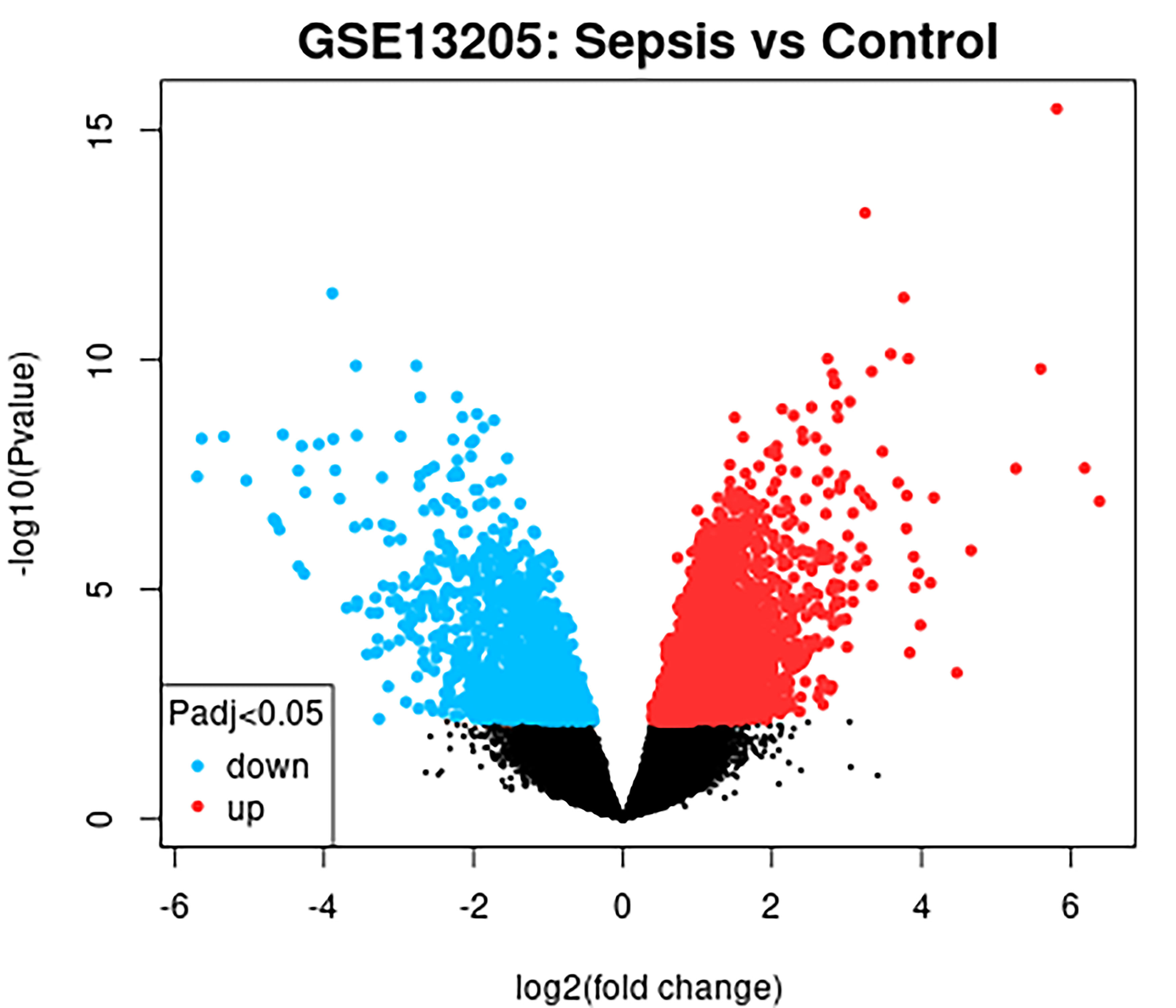
Using GEO2R analysis with the criteria of P<0.05 and |LogFC| > 1, a total of 2232 DEGs were identified in the GSE13205 dataset, comprising 1469 upregulated and 763 downregulated genes. The volcano plot visualised these DEGs, with red and blue dots representing upregulated and downregulated genes, respectively (Fig. 1).
Fig. 1. Analysis of DEGs in sepsis. Volcano plot of gene expression in septic patients from the GSE13205 dataset.
Identification of PRDEGs
Pyroptosis-related genes were obtained from the “GeneCards” database and intersected with DEGs in GSE13205 to identify PRDEGs. As shown in the Venn diagram, after removing duplicate gene symbols, 1667 genes on the left were unique to GSE13205, 39 genes in the middle were PRDEGs, and 716 genes on the right were uniquely related to pyroptosis (Fig. 2A). Visualisation of PRDEGs was further done using a heatmap (Fig. 2B), with 32 genes upregulated and 7 genes downregulated (Supplementary Table S1).
Fig. 2. Pyroptosis-related genes and Venn analysis. (A) Intersection of DEGs related to pyroptosis. (B) Heatmap of pyroptosis-related genes.
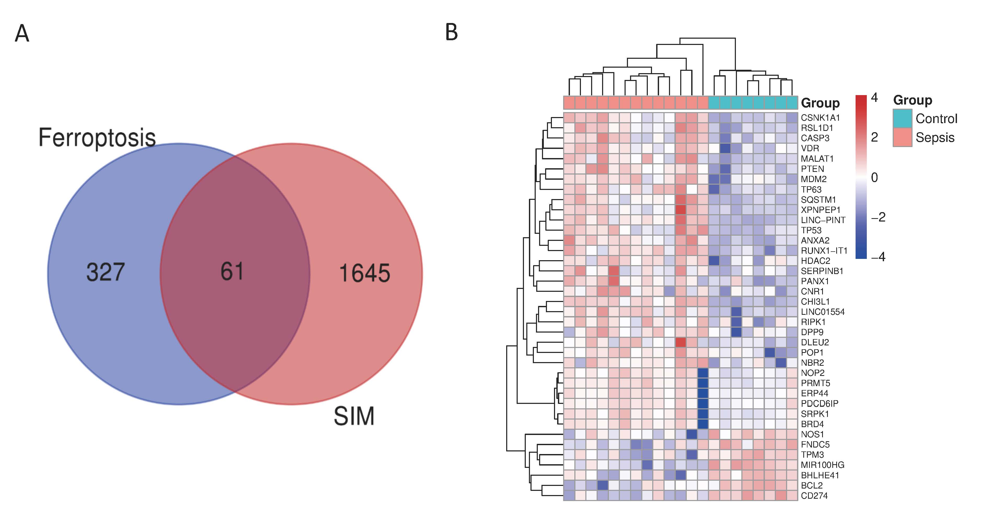
SIM: sepsis-induced myopathy
GO and KEGG enrichment analysis of PRDEGs
Metascape database was utilised for GO and KEGG enrichment analysis of PRDEGs and visualisation of the results. In the MF enrichment analysis, PRDEGs were primarily enriched in functions related to enzyme binding, p53 binding, ubiquitin-protein binding, calcium-dependent protein binding, ubiquitin-like protein binding and protein serine/threonine kinase activity. In the BP enrichment, PRDEGs were primarily associated with the positive regulation of cell apoptosis, cell death, programmed cell death, regulation of the p53 signalling pathway, circadian rhythms and cellular responses to oxygen levels, ischaemia and chemical stimuli. CC enrichment revealed associations with myelin sheath, membrane microdomains, membrane vesicles, promyelocytic leukaemia (PML) bodies, transcription factor complexes, nuclear matrix and various vesicles (Fig. 3A). KEGG enrichment analysis highlighted pathways such as the p53 signalling pathway, microRNAs in cancer and associated pathways, apoptosis and endocrine resistance (Fig. 3B).
Fig. 3. Enrichment analysis of PRDEGs using the metascape database. (A) GO enrichment analysis, including biological processes and cellular components pathways. (B) Kyoto Encyclopedia of Genes and Genomes enrichment analysis. 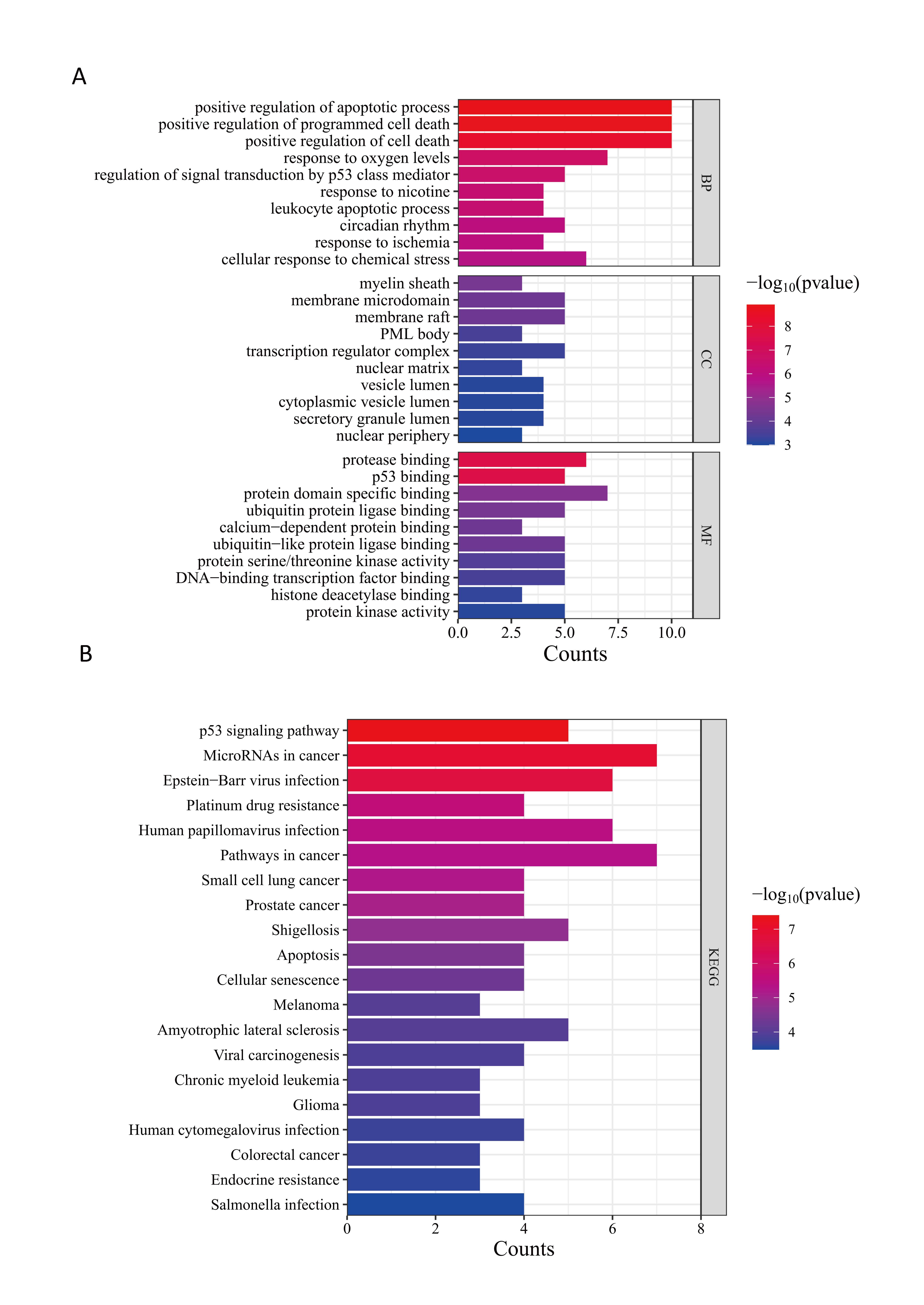
GO: Gene Ontology; PML: promyelocytic leukaemia; PRDEGs: pyroptosis-related differentially expressed genes
Construction of PPI network for PRDEGs and identification of hub genes
A PPI network for PRDEGs was constructed using the STRING database with an interaction score threshold of >0.4, resulting in 25 nodes and 140 edges. Among the 39 PRDEGs, 14 genes had no correlations with other genes and did not form molecular networks. These were visualised using Cytoscape software (Cytoscape Consortium, San Diego, CA, US), and the most important module in the PPI network was identified using the MCODE plugin (Figs. 4A, C). Subsequently, hub genes were identified using the CytoHubba plugin in Cytoscape, with the top 10 genes ranked by the MCC algorithm (Supplementary Table S2). Functional enrichment analysis of hub genes using the Metascape database revealed enriched functions and pathways, including TP53, PTEN, BCL2, CASP3, MDM2, BRD4, HDAC2, CD274, SQSTM1 and CSNK1A1 (Figs. 4B, D).
Fig. 4. (A, C) Construction of the protein–protein interaction network using the STRING database. (B, D) Identification of hub genes using the CytoHubba plugin in Cytoscape.

Internal validation of hub gene expression differences
Expression matrices of hub genes were obtained from the GEO database, and their expression differences were visualised using violin plots. Statistical analysis revealed significant upregulation of TP53, PTEN, CASP3, MDM2, BRD4, HDAC2, SQSTM1 and CSNK1A1 in the skeletal muscle of sepsis patients compared to the control group, while BCL2 and CD274 exhibited significant downregulation (Fig. 5).
Fig. 5. Expression matrix of hub genes obtained from the GEO database.
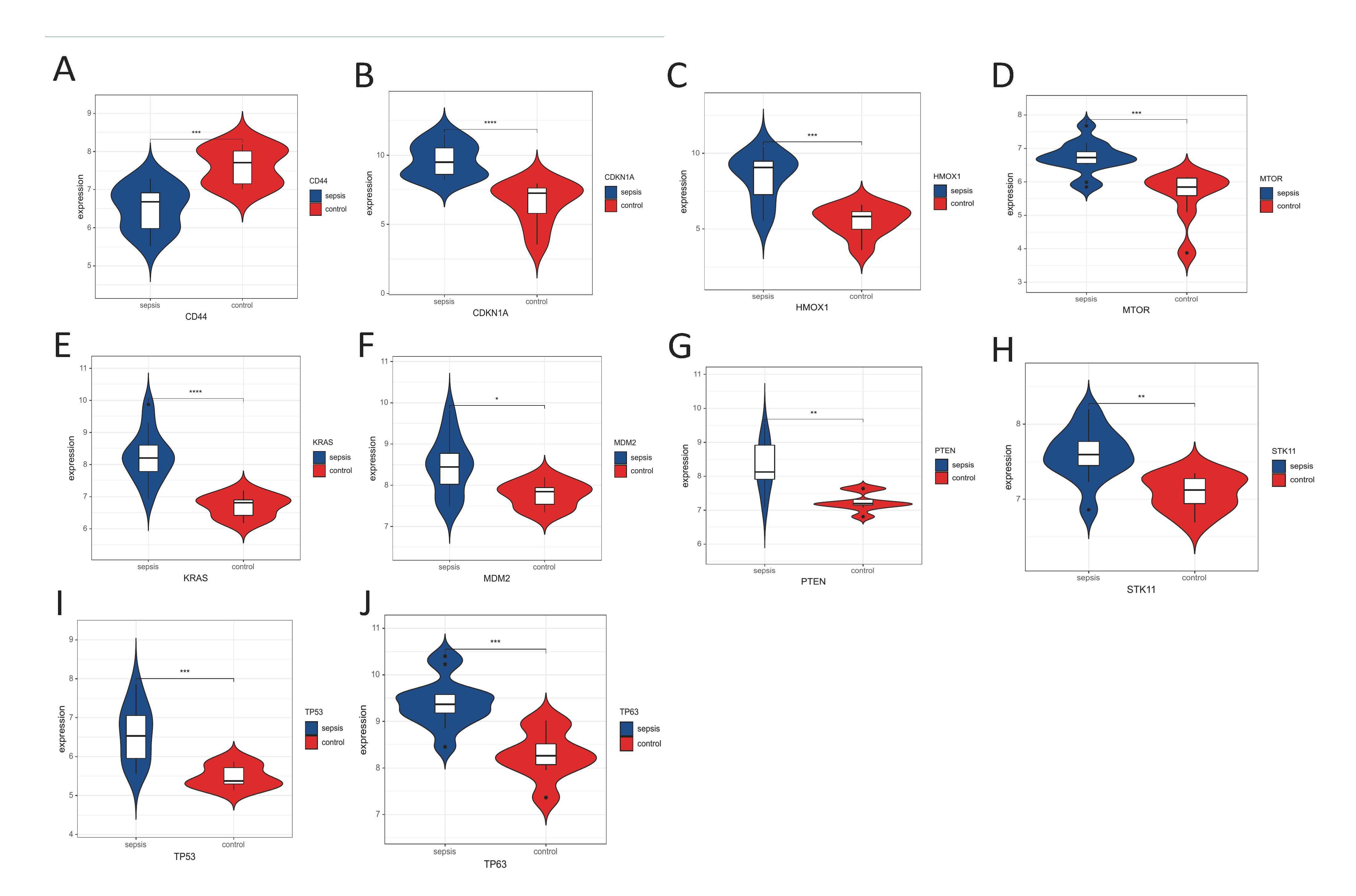
GEO: Gene Expression Omnibus
*Comparison with the control group, P<0.05
**Comparison with the control group, P<0.01
***Comparison with the control group, P<0.001
CLP-induced sepsis significantly alters the mRNA expression of key genes in mouse skeletal muscle
To further confirm the impact of CLP-induced sepsis on gene expression in skeletal muscle, the mRNA levels of 10 key genes in skeletal muscle tissues from sham and CLP mice were examined using qRT-PCR. The results revealed that the mRNA expression levels of TP53, PTEN, BCL2, CASP3, MDM2, BRD4, HDAC2, SQSTM1 and CSNK1A1 were significantly elevated in the CLP group compared with the sham group (Figs. 6A–F, H–J; P<0.001), suggesting that CLP stimulation markedly activates pathways related to cellular stress, apoptosis and epigenetic regulation. Notably, the expression of the immune checkpoint molecule CD274 (PD-L1) was significantly decreased in the CLP group (Fig. 6G; P<0.001), which may be associated with the development of an immunosuppressive state. These findings indicate that CLP-induced sepsis profoundly disturbs the expression of multiple critical genes in skeletal muscle, involving diverse BP such as cell death, inflammatory regulation and signal transduction.
Fig. 6. qRT-PCR was performed to assess the mRNA expression levels of multiple genes associated with programmed cell death, inflammation, and signal regulation in skeletal muscle tissues from sham and CLP groups. The analysed genes included (A) TP53, (B) PTEN, (C) BCL2, (D) CASP3, (E) MDM2, (F) BRD4, (G) CD274, (H) HDAC2, (I) SQSTM1 and (J) CSNK1A1.
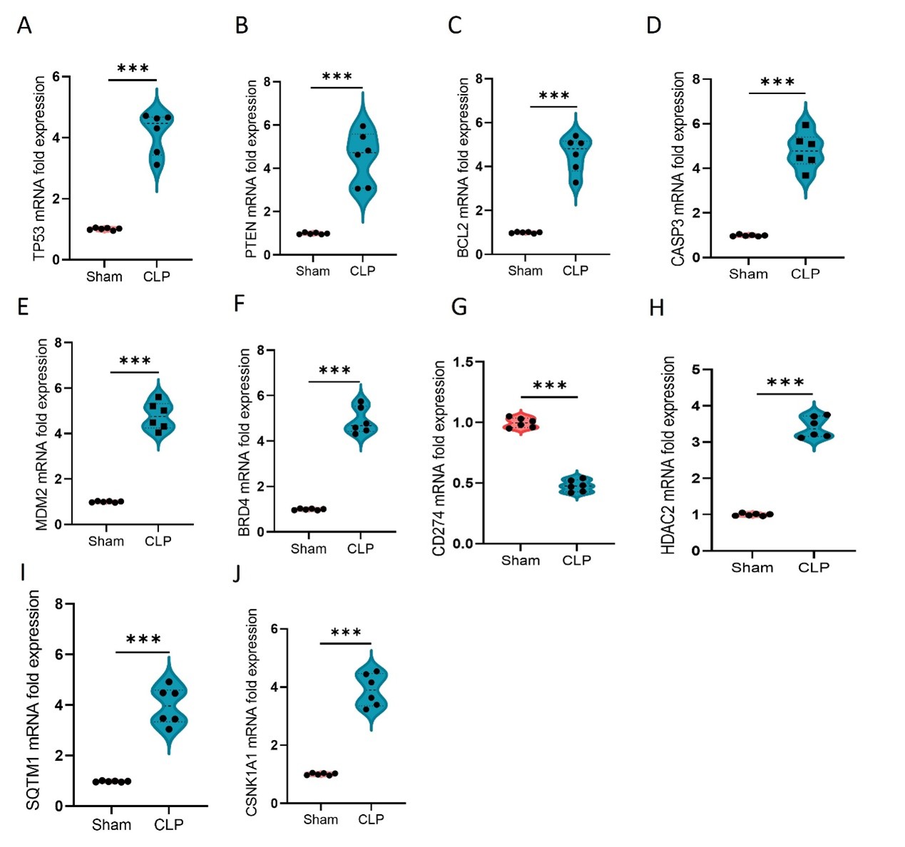
CLP: cecal ligation and puncture; qRT-PCRL: real-time quantitative polymerase chain reaction
Note: Except for CD274, all other genes were significantly upregulated in the CLP group. Each dot represents an individual mouse (n=6–9). Data are presented as violin plots, with the centre point indicating the median.
***P<0.001, analysed by conducting the non-parametric Mann–Whitney test.
Screening for potential pharmacological targets
The screening results for potential pharmacological targets were obtained from CMap based on connectivity scores. The top 10 molecular drugs with therapeutic potential for SIM were identified as Quinpirole, Zolpidem, PTB1, CG-930, 9-methyl-5H-6-thia-4,5-diaza-chrysene-6,6-dioxide, VER-155008, Chromanol, Anagrelide, Tipifarnib-P2 and Benproperine (Supplementary Table S3). These drugs hold promise as potential treatments for pyroptosis-induced SIM.
DISCUSSION
Apoptosis is a non-inflammatory cell death process, typically occurring during cellular self-repair and the removal of damaged cells. In contrast, pyroptosis is an inflammatory cell death process triggered by infection or inflammation, accompanied by a significant inflammatory response.8 Previous studies have indicated that the activation of the pyroptosis pathway plays a crucial role in the progression of SIM.8,16 Preclinical research has identified several related targets that can mitigate muscle wasting.16-18 Therefore, inhibiting the inflammatory signalling pathways associated with pyroptosis holds promise as a therapeutic strategy to improve the prognosis of SIM. However, the role of pyroptosis-related genes in SIM remains incompletely understood, and this study aimed to clarify this role.
To elucidate the expression of pyroptosis-related genes in SIM, the GSE13205 dataset was utilised, and 39 genes associated with pyroptosis were identified. Among these, 32 genes were upregulated and 7 genes were downregulated in SIM compared to normal tissues. This suggests that pyroptosis-related genes may be involved in the pathological processes of SIM. To gain deeper insights into the roles of these pyroptosis-related genes in SIM, the GO enrichment analysis was conducted. The enriched MF included positive regulation of apoptosis process, positive regulation of programmed cell death, positive regulation of cell death, response to oxygen levels, regulation of signal transduction by p53 class mediator, response to nicotine, leukocyte apoptotic process, circadian rhythm, response to ischaemia and cellular response to chemical stress. These findings indicate a predominant involvement of pyroptosis-related genes in positively regulating cell death or apoptosis. It has been reported that programmed cell death, including autophagy, apoptosis, pyroptosis and necroptosis, plays a significant role in SIM.19,20 Studies have shown that sepsis induces muscle fibre apoptosis, leading to skeletal muscle atrophy.19 The activation of the NLRP3 inflammasome pyroptosis pathway is also considered a potential mechanism and therapeutic target in SIM, but the exact contribution of this pathway in SIM remains to be fully validated experimentally.16,20,21 Sepsis-induced limb muscle fibre atrophy is associated with enhanced activity of the proteasome and autophagy protein degradation pathways, triggered by the inhibition of protein kinase B (AKT) and mammalian target of rapamycin complex 1 and the activation of the adenosine monophosphate-activated protein kinase pathway.22 Oxidative stress, such as reactive oxygen species is another critical initiating factor and therapeutic target for skeletal muscle atrophy in sepsis.21,23 It is also a key mechanism through which some drugs exert their therapeutic effects on SIM.24 In response to cellular and extracellular stimuli and stress, p53 acts as a transcription factor regulating the expression of downstream genes to help cells/organisms resist these stimuli. Although some studies have identified p53 as a regulator of pyroptosis,25 its role in SIM remains unclear. Interestingly, circadian rhythm is associated with the activation of the NLRP3 inflammasome.26 Research has found that continuous light exacerbates skeletal muscle loss in endotoxemia rats, which is related to changes in the hypothalamic circadian clock and SIRT1.27 The MF highlighted in GO enrichment provides crucial information about the biological roles that these genes may play in SIM, offering insights into the potential role of pyroptosis in SIM.
The impact of PRDEGs in SIM spans multiple biological pathways. KEGG pathway analysis revealed that PRDEGs in SIM are mainly involved in pathways such as the p53 signalling pathway, microRNAs in cancer, Epstein-Barr virus infection, platinum drug resistance, human papillomavirus infection, pathways in cancer, small cell lung cancer, prostate cancer, Shigellosis and apoptosis. Apart from the p53 signalling pathway and apoptosis, these genes are associated with several cancer-related pathways. Cellular pyroptosis is involved in tumour proliferation, invasion and metastasis and is a therapeutic target in numerous cancers.28 Given the similarities in pathological manifestations and characteristics between SIM and cachexia induced by tumours, these enriched pathways could provide novel insights for treatment strategies in SIM. However, experimental validation of these pathways in SIM is required to confirm their role in the disease.29,30 These enriched pathways can provide valuable insights and guidance for the treatment of SIM. A PPI network of PRDEGs was also constructed using STRING, and 10 core genes in pyroptosis associated with SIM were identified: TP53, PTEN, MDM2, SQSTM1, CASP3, CSNK1A1, BRD4, BCL2, HDAC2, CD274, which were further validated using the CytoHubba plugin in Cytoscape. Further investigation into the functions and interactions of these genes, particularly through in vitro and in vivo experiments, will provide a better understanding of their contribution to SIM and their potential as therapeutic targets. These key genes related to pyroptosis may serve as potential therapeutic targets that require further research.
In addition, the authors screened for potential pharmacological targets by downloading results from the CMap database and ranking and filtering them based on drug connectivity scores. The top 10 molecular drugs identified were Quinpirole, Zolpidem, PTB1, CG-930, 9-methyl-5H-6-thia-4,5-diaza-chrysene-6,6-dioxide, VER-155008, Chromanol, Anagrelide, Tipifarnib-P2 and Benproperine. However, these drugs currently lack preclinical validation in SIM, and their effectiveness in this context needs to be further investigated. Future experimental studies should validate these drugs’ potential impact on the pyroptosis-related genes identified in this study.
This study has limitations. First, it lacks relevant clinical information about patient characteristics, such as age, sex and disease duration, which could influence gene expression. Additionally, a larger sample size of SIM patients may be considered to validate the stability of the results of this study. Furthermore, future studies should include in vitro and in vivo experiments to validate the therapeutic potential of the identified compounds.
CONCLUSION
In summary, the pathogenesis of SIM involves multiple BP and signalling pathways. This study has shown significant changes in the expression of pyroptosis-related genes in SIM, suggesting a potential role of pyroptosis in the pathogenesis of this disease. Based on the results, these pyroptosis-related genes may serve as potential pharmacological targets. Interventions targeting these genes could provide a promising approach for regulating the development of SIM. Moreover, this study provides a foundation for further exploration of the role of pyroptosis in sepsis and offers new directions for disease treatment research.
Table S1. 39 PRDEGs.
Table S2. Top 10 hub genes identified by CytoHubba.
Table S3. Top 10 small molecular compounds provided by CMap to PRDEGs.
REFERENCES
- Singer M, Deutschman CS, Seymour CW, et al. The Third International Consensus Definitions for Sepsis and Septic Shock (Sepsis-3). JAMA 2016;315:801-10.
- Gardner AK, Ghita GL, Wang Z, et al. The Development of Chronic Critical Illness Determines Physical Function, Quality of Life, and Long-Term Survival Among Early Survivors of Sepsis in Surgical ICUs. Crit Care Med 2019;47:566-73.
- Callahan LA, Supinski GS. Sepsis-induced myopathy. Crit Care Med 2009;37:S354-67.
- Dres M, Goligher EC, Heunks LMA, et al. Critical illness-associated diaphragm weakness. Intensive Care Med 2017;43:1441-52.
- Liu Y, Xu L, Yang Z, et al. Gut-muscle axis and sepsis-induced myopathy: The potential role of gut microbiota. Biomed Pharmacother 2023;163:114837.
- Mankowski RT, Laitano O, Clanton TL, et al. Pathophysiology and Treatment Strategies of Acute Myopathy and Muscle Wasting after Sepsis. J Clin Med 2021;10:1874.
- Oikonomou E, Paraskevas T, Velissaris D. Sepsis and the muscle tissue. A narrative review. Rom J Intern Med 2021;59:218-26.
- Liu Y, Wang D, Li T, et al. The role of NLRP3 inflammasome in inflammation-related skeletal muscle atrophy. Front Immunol 2022;13:1035709.
- Zheng X, Chen W, Gong F, et al. The Role and Mechanism of Pyroptosis and Potential Therapeutic Targets in Sepsis: A Review. Front Immunol 2021;12:711939.
- Wang Y, Liu Y, Liu Q, et al. Caspase-1-Dependent Pyroptosis of Peripheral Blood Mononuclear Cells Is Associated with the Severity and Mortality of Septic Patients. Biomed Res Int 2020;9152140.
- Wen R, Liu YP, Tong XX, et al. Molecular mechanisms and functions of pyroptosis in sepsis and sepsis-associated organ dysfunction. Front Cell Infect Microbiol 2022;12:962139.
- Gou X, Xu W, Liu Y, et al. IL-6 Prevents Lung Macrophage Death and Lung Inflammation Injury by Inhibiting GSDME- and GSDMD-Mediated Pyroptosis during Pneumococcal Pneumosepsis. Microbiol Spectr 2022;10:e0204921.
- Jin H, Xie W, He M, et al. Pyroptosis and Sarcopenia: Frontier Perspective of Disease Mechanism. Cells 2022;11:1078.
- Jimenez-Gutierrez GE, Martinez-Gomez LE, Martinez-Armenta C, et al. Molecular Mechanisms of Inflammation in Sarcopenia: Diagnosis and Therapeutic Update. Cells 2022;11:2359.
- Wang L, Jiao XF, Wu C, et al. Trimetazidine attenuates dexamethasone-induced muscle atrophy via inhibiting NLRP3/GSDMD pathway-mediated pyroptosis. Cell Death Discov 2021;7:251.
- Huang N, Kny M, Riediger F, et al. Deletion of Nlrp3 protects from inflammation-induced skeletal muscle atrophy. Intensive Care Med Exp 2017;5:3.
- Liu Y, Zhang Y, Wang C, et al. Inhibition of DDX3X alleviates persistent inflammation, immune suppression and catabolism syndrome in a septic mice model. Int Immunopharmacol 2023;117:109779.
- Wang C, Liu Y, Zhang Y, et al. Targeting NAT10 protects against sepsis-induced skeletal muscle atrophy by inhibiting ROS/NLRP3. Life Sci 2023;121948.
- Cao YY, Wang Z, Yu T, et al. Sepsis induces muscle atrophy by inhibiting proliferation and promoting apoptosis via PLK1-AKT signalling. J Cell Mol Med 2021;25:9724-39.
- Liu Y, Wang D, Li T, et al. The role of NLRP3 inflammasome in inflammation-related skeletal muscle atrophy. Front Immunol 2022;13:1035709.
- Wang C, Liu Y, Zhang Y, et al. Targeting NAT10 protects against sepsis-induced skeletal muscle atrophy by inhibiting ROS/NLRP3. Life Sci 2023;330:121948.
- Stana F, Vujovic M, Mayaki D, et al. Differential Regulation of the Autophagy and Proteasome Pathways in Skeletal Muscles in Sepsis. Crit Care Med 2017;45:e971-9.
- Yang B, Yang X, Sun X, et al. IL-6 Deficiency Attenuates Skeletal Muscle Atrophy by Inhibiting Mitochondrial ROS Production through the Upregulation of PGC-1alpha in Septic Mice. Oxid Med Cell Longev 2022;9148246.
- Yu X, Han W, Wang C, et al. Upregulation of Heme Oxygenase-1 by Hemin Alleviates Sepsis-Induced Muscle Wasting in Mice. Oxid Med Cell Longev 2018;2018:8927104.
- Huang Y, Yong P, Dickey D, et al. Inflammasome Activation and Pyroptosis via a Lipid-regulated SIRT1-p53-ASC Axis in Macrophages From Male Mice and Humans. Endocrinology 2022;163:bqac014.
- Adamiak M, Ciechanowicz A, Skoda M, et al. Novel Evidence that Purinergic Signaling – Nlrp3 Inflammasome Axis Regulates Circadian Rhythm of Hematopoietic Stem/Progenitor Cells Circulation in Peripheral Blood. Stem Cell Rev Rep 2020;16:335-43.
- Chen Y, Cheng M, Su T, et al. Constant light exposure aggravates POMC-mediated muscle wasting associated with hypothalamic alteration of circadian clock and SIRT1 in endotoxemia rats. Biochem Biophys Res Commun 2019;508:811-7.
- Ruan J, Wang S, Wang J. Mechanism and regulation of pyroptosis-mediated in cancer cell death. Chem Biol Interact 2020;323:109052.
- Lee JH, Kim SW, Kim JH, et al. Lithium Chloride Protects against Sepsis-Induced Skeletal Muscle Atrophy and Cancer Cachexia. Cells 2021;10:1017.
- Hall DT, Ma JF, Marco SD, et al. Inducible nitric oxide synthase (iNOS) in muscle wasting syndrome, sarcopenia, and cachexia. Aging (Albany NY) 2011;3:702-15.
All animal experiments were conducted in accordance with the ARRIVE guidelines. The experimental protocols were approved by the Animal Ethics Committee of Tongji Hospital, Tongji Medical College, Huazhong University of Science and Technology (TJH-24-06-005). All procedures were carried out in compliance with the UK Animals (Scientific Procedures) Act, 1986 and associated guidelines, the EU Directive 2010/63/EU for animal experiments.
This study was supported by grants from Hubei Provincial Natural Science Foundation of China (2023AFB216). The authors declare there are no affiliations with or involvement in any organisation or entity with any financial interest in the subject matter or materials discussed in this manuscript. The authors also declare there is no conflict of interest.
Prof Yukun Liu, Department of Plastic and Aesthetic Surgery, Tongji Hospital, Tongji Medical College, Huazhong University of Science and Technology, Wuhan, China 430030. Email: [email protected]; Prof Yuchang Wang, Division of Trauma Surgery, Emergency Surgery & Surgical Critical, Tongji Hospital, Tongji Medical College, Huazhong University of Science and Technology, Wuhan, China 430030. Email: [email protected].
